Anti-Retinoic Acid Receptor beta抗体
参阅全部 Retinoic Acid Receptor beta 一抗
兔多克隆抗体to Retinoic Acid Receptor beta
Rabbit
This antibody shows slight cross-reactivity to RAR alpha but does not detect RAR gamma.
适用于: ICC/IF, WBmore details
与反应: Mouse, Human
Synthetic peptide corresponding to Mouse Retinoic Acid Receptor beta aa 400-500.
WB: SH-SY5Y cell extract ; HEK-293 ICC/IF: SH-SY5Y
The Life Science industry has been in the grips of a reproducibility crisis for a number of years. Abcam is leading the way in addressing this with our range of recombinant monoclonal antibodies and knockout edited cell lines for gold-standard validation. Please check that this product meets your needs before purchasing.
If you have any questions, special requirements or concerns, please send us an inquiry and/or contact our Support team ahead of purchase. Recommended alternatives for this product can be found below, along with publications, customer reviews and Q&As
Liquid
Shipped at 4°C. Store at +4°C short term (1-2 weeks). Upon delivery aliquot. Store at -20°C or -80°C. Avoid freeze / thaw cycle.
Preservative: 0.05% Sodium azide
Constituent: 99% PBS
Whole antiserum
多克隆
IgG
Abpromise™承诺保证使用ab5792于以下的经测试应用
“应用说明”部分 下显示的仅为推荐的起始稀释度;实际最佳的稀释度/浓度应由使用者检定。
| 应用 | Ab评论 | 说明 |
|---|---|---|
| ICC/IF | 1/100. | |
| WB | 1/1000. Detects a band of approximately 52 kDa (predicted molecular weight: 53 kDa). |
Entrez Gene: 5915 Human
Entrez Gene: 218772 Mouse
Omim: 180220 Human
SwissProt: P10826 Human
SwissProt: P22605 Mouse
Unigene: 543218 Human
Unigene: 581530 Human
Unigene: 654490 Human
Unigene: 733004 Human
Unigene: 259318 Mouse
HAP antibody
HBV-activated protein antibody
NR1B2 antibody
Nuclear receptor subfamily 1 group B member 2 antibody
RAR B antibody
RAR beta antibody
RAR epsilon antibody
RAR-beta antibody
RAR-epsilon antibody
RARB antibody
RARB_HUMAN antibody
Retinoic acid receptor beta 2 antibody
Retinoic acid receptor beta 4 antibody
Retinoic acid receptor beta 5 antibody
Retinoic acid receptor beta antibody
Retinoic acid receptor beta polypeptide antibody
RRB2 antibody

Western blot - Anti-Retinoic Acid Receptor beta antibody (ab5792)
All lanes : Anti-Retinoic Acid Receptor beta antibody (ab5792) at 1 µg/ml
Lane 1 : SH-SY5Y (Human neuroblastoma cell line from bone marrow) whole cell lysate
Lane 2 : HEK-293 (Human epithelial cell line from embryonic kidney) whole cell lysate
Lane 3 : PANC-1 (Human pancreatic epithelial cancinoma cell line) whole cell lysate
Lane 4 : OVCAR-3 (Human ovary adenocarcinoma cell line) whole cell lysate
Lane 5 : BeWo (human placenta choriocarcinoma cell line) whole cell lysate
Lysates/proteins at 30 µg per lane.
Secondary
All lanes : Goat anti-Rabbit IgG (H+L), Superclonal™ Recombinant Secondary Antibody, HRP at 1/4000 dilution
Predicted band size: 53 kDa
Additional bands at: ~58.50 kDa. We are unsure as to the identity of these extra bands.
Detection: chemiluminescence
Western blot demonstrating antibody specificity by detection of differential basal expression of the target across cell lines owing to their inherent genetic constitution. The expression was observed in SH-SY5Y and HEK-293 and not seen in PANC-1, OVCAR-3 and BeWo using ab5792.
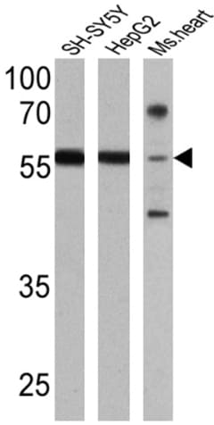
Western blot - Anti-Retinoic Acid Receptor beta antibody (ab5792)
All lanes : Anti-Retinoic Acid Receptor beta antibody (ab5792) at 1/1000 dilution
Lane 1 : SH-SY5Y cell lysate
Lane 2 : HepG2 cell lysate
Lane 3 : Mouse heart cell lysate
Lysates/proteins at 25 µg per lane.
Predicted band size: 53 kDa
Observed band size: 58 kDawhy is the actual band size different from the predicted?

Immunocytochemistry/ Immunofluorescence - Anti-Retinoic Acid Receptor beta antibody (ab5792)
Immunofluorescent analysis of SH-SY5Y (Human neuroblastoma cell line from bone marrow) whole cell lysate cells on 70% confluent log phase labeling Retinoic Acid Receptor beta. The cells were fixed with 4% paraformaldehyde for 10 minutes, permeabilized with 0.1% Triton™ X-100 for 10 minutes, and blocked with 2% BSA for 10 minutes at room temperature. The cells were labeled with ab5792 at 1/100 dilution in 0.1% BSA and incubated overnight at 4°C and then labeled with Goat anti-Rabbit IgG (H+L) secondary antibody, Alexa Fluor® 488 conjugate at 1/2000 dilution for 45 minutes at room temperature (Panel a: green). Nuclei (Panel b: blue) were stained with DAPI. F-actin (Panel c: red) was stained with Alexa Fluor® 555 Rhodamine Phalloidin (1/300 dilution). Panel d is a merged image showing nuclear and cytoplasmic localization. Panel e represents BeWo (human placenta choriocarcinoma cell line) having no expression of Retinoic Acid Receptor beta. The images were captured at 60X magnification.


