Anti-Caveolin-1抗体- Caveolae Marker
参阅全部 Caveolin-1 一抗
兔多克隆抗体to Caveolin-1 - Caveolae Marker
Rabbit
适用于: WB, ICC/IF, IPmore details
与反应: Mouse, Rat, Human
预测可用于: Sheep, Rabbit, Horse, Cow, Cat, Pig, Chimpanzee, Gorilla, African green monkey, African bush elephant![]()
Synthetic peptide corresponding to Human Caveolin-1 aa 1-100.
Database link: Q03135
WB: Human lung, heart and spleen tissue lysates; Mouse heart and lung tissue lysates; Rat heart protein extract; PANC-1, U-87 MG, A549, HeLa, PC-3, U-2 OS, C2C12 and A431 cell lysates. ICC/IF: A-375, HeLa and NIH/3T3 cells; Rat astrocytes.
This antibody can be used as a marker for lipid raft fractions.
The Life Science industry has been in the grips of a reproducibility crisis for a number of years. Abcam is leading the way in addressing this with our range of recombinant monoclonal antibodies and knockout edited cell lines for gold-standard validation. Please check that this product meets your needs before purchasing.
If you have any questions, special requirements or concerns, please send us an inquiry and/or contact our Support team ahead of purchase. Recommended alternatives for this product can be found below, along with publications, customer reviews and Q&As
Abpromise™承诺保证使用ab2910于以下的经测试应用
“应用说明”部分 下显示的仅为推荐的起始稀释度;实际最佳的稀释度/浓度应由使用者检定。
| 应用 | Ab评论 | 说明 |
|---|---|---|
| WB | (21) | Use a concentration of 1 - 2 µg/ml. Detects a band of approximately 22 kDa (predicted molecular weight: 20 kDa). |
| ICC/IF | (15) | Use at an assay dependent concentration. |
| IP | (9) | Use at an assay dependent concentration. Recommended dilution: 5 µg |
Entrez Gene: 463670 Chimpanzee
Entrez Gene: 101125469 Gorilla
Entrez Gene: 857 Human
Entrez Gene: 12389 Mouse
Entrez Gene: 100008837 Rabbit
Entrez Gene: 445468 Sheep
Omim: 601047 Human
SwissProt: Q2QLF2 Chimpanzee
SwissProt: Q2IBF0 Gorilla
SwissProt: Q2QLB0 Horse
SwissProt: Q03135 Human
SwissProt: P49817 Mouse
SwissProt: Q09YN6 Rabbit
SwissProt: Q6B3Y2 Sheep
Unigene: 74034 Human
Unigene: 28278 Mouse
Unigene: 470537 Mouse
Unigene: 22518 Rat
BSCL3 antibody
CAV antibody
CAV1 antibody
CAV1_HUMAN antibody
caveolae protein, 22 kD antibody
caveolin 1 alpha isoform antibody
caveolin 1 beta isoform antibody
Caveolin 1 caveolae protein 22kDa antibody
Caveolin-1 antibody
Caveolin1 antibody
cell growth-inhibiting protein 32 antibody
CGL3 antibody
LCCNS antibody
MSTP085 antibody
OTTHUMP00000025031 antibody
PPH3 antibody
VIP 21 antibody
VIP21 antibody

Western blot - Anti-Caveolin-1 antibody - Caveolae Marker (ab2910)
All lanes : Anti-Caveolin-1 antibody - Caveolae Marker (ab2910) at 1.5 µg/ml
Lane 1 : Human lung
Lane 2 : Human heart
Lane 3 : Human spleen
Lysates/proteins at 20 µg per lane.
Secondary
All lanes : Alexa Fluor anti-rabbit at 1/5000 dilution
Predicted band size: 20 kDa
Observed band size: 20 kDa

Immunocytochemistry/ Immunofluorescence - Anti-Caveolin-1 antibody - Caveolae Marker (ab2910)
ab2910 staining Caveolin-1 in wild-type HeLa cells (top panel) and CAV1 knockout HeLa cells (ab255371) (bottom panel). The cells were fixed with 100% methanol (5 min) then permeabilized with 0.1% Triton X-100 for 5 minutes and then blocked with 1% BSA/10% normal goat serum/0.3M glycine in 0.1% PBS-Tween for 1h. The cells were then incubated with ab2910 at 1/500 dilution and ab7291 (Mouse monoclonal to alpha Tubulin) at 1/1000 dilution overnight at 4°C followed by a further incubation at room temperature for 1h with a goat secondary antibody to rabbit IgG (Alexa Fluor® 488) (ab150081) at 2 μg/ml (shown in green) and a goat secondary antibody to mouse IgG (Alexa Fluor® 594) (ab150120) at 2 μg/ml (shown in red). Nuclear DNA was labelled in blue with DAPI.
Image was taken with a confocal microscope (Leica-Microsystems TCS SP8).

Immunoprecipitation - Anti-Caveolin-1 antibody - Caveolae Marker (ab2910)
Caveolin 1 was immunoprecipitated using 5 µg of ab2910 from lysate of Mouse Heart (Lane 3) using the Dynabeads® Protein A Immunoprecipitation Kit. Normal Rabbit IgG was used as a Isotype control (Lane 2). 10 % input represents the cell extract used for immunoprecipitation (Lane 1). Western blot analysis was performed using Caveolin 1 Rabbit Polyclonal Antibody and Goat anti-Rabbit IgG (H+L) Superclonal™ Secondary Antibody, HRP conjugate. Chemiluminescent detection was performed using Pierce™ ECL Western Blotting Substrate.
All lanes :
Lane 1 : Input control
Lane 2 : Isotype control
Lane 3 : IP elute
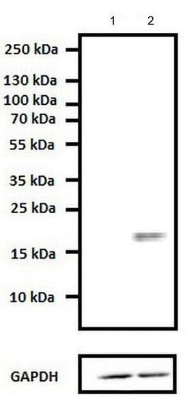
Western blot - Anti-Caveolin-1 antibody - Caveolae Marker (ab2910)
All lanes : Anti-Caveolin-1 antibody - Caveolae Marker (ab2910) at 1 µg/ml
Lane 1 : CAV1 knockout HeLa cell lysate
Lane 2 : Wild-type HeLa cell lysate
Predicted band size: 20 kDa
The specificity of ab2910 was demonstrated by CRISPR targeted CAV1 knockout in HeLa cells. Western blot analysis of whole cell lysates using this antibody showed no detection of caveolin 1 protein expression in knockout cells compared to the protein detected at ~22kDa in wild-type HeLa cells.

Western blot - Anti-Caveolin-1 antibody - Caveolae Marker (ab2910)
All lanes : Anti-Caveolin-1 antibody - Caveolae Marker (ab2910) at 1 µg/ml
Lane 1 : PANC-1 (Human pancreatic epithelial cancinoma cell line) whole cell lysate
Lane 2 : U-87 MG (Human glioblastoma-astrocytoma epithelial cell line) whole cell lysate
Lane 3 : A549 (Human lung carcinoma cell line) whole cell lysate
Lane 4 : HeLa (Human cervix adenocarcinoma epithelial cell) whole cell lysate
Lane 5 : PC-3 (Human prostate adenocarcinoma cell line) whole cell lysate
Lane 6 : U-2 OS (Human bone osteosarcoma epithelial cell line) whole cell lysate
Lane 7 : Mouse heart tissue lysate
Lane 8 : Rat heart tissue lysate
Lane 9 : C2C12 (Mouse myoblast cell line) whole cell lysate
Lane 10 : Mouse lung tissue lysate
Lane 11 : A-431 (Human epidermoid carcinoma cell line) whole cell lysate
Lysates/proteins at 20 µg per lane.
Predicted band size: 20 kDa
Western blot analysis was performed on whole cell extracts (20 µg lysate). The blots were probed with Anti-ab2910 (1-2 µg/mL) and detected by chemiluminescence using Goat anti-Rabbit IgG (H+L) Superclonal™ Secondary Antibody, HRP conjugate. A 17 kDa band corresponding to Caveolin-1 was observed across cell lines and tissues tested. Known quantity of protein samples were electrophoresed using Novex® NuPAGE® 12 % Bis-Tris gel, XCell SureLock™ Electrophoresis System and Novex® Sharp Pre-Stained Protein Standard. Resolved proteins were then transferred onto a nitrocellulose membrane with iBlot® 2 Dry Blotting System. The membrane was probed with the relevant primary and secondary Antibody following blocking with 5 % skimmed milk. Chemiluminescent detection was performed using Pierce™ ECL Western Blotting Substrate.

Western blot - Anti-Caveolin-1 antibody - Caveolae Marker (ab2910)
Anti-Caveolin-1 antibody - Caveolae Marker (ab2910) at 2 µg/ml + Rat heart protein extract
Predicted band size: 20 kDa

Immunocytochemistry/ Immunofluorescence - Anti-Caveolin-1 antibody - Caveolae Marker (ab2910)
Immunofluorescence analysis of Caveolin 1 was done on 70% confluent log phase A-375 cells. The cells were fixed with 4% paraformaldehyde for 15 minutes, permeabilized with 0.25% Triton™ X-100 for 10 minutes, and blocked with 5% BSA for 1 hour at room temperature. The cells were labeled with ab2910 at 1 µg/mL in 1% BSA and incubated for 3 hours at room temperature and then labeled with Goat anti-Rabbit IgG (H+L) Superclonal™ Secondary Antibody, Alexa Fluor® 488 conjugate at a dilution of 1:2000 for 45 minutes at room temperature (Panel a: green). Nuclei (Panel b: blue) were stained with SlowFade® Gold Antifade Mountant with DAPI. F-actin (Panel c: red) was stained with Alexa Fluor® 555 Rhodamine Phalloidin. Panel d is a merged image showing cytoplasmic localization. Panel e is a no primary antibody control. The images were captured at 60X magnification.

Immunocytochemistry/ Immunofluorescence - Anti-Caveolin-1 antibody - Caveolae Marker (ab2910)
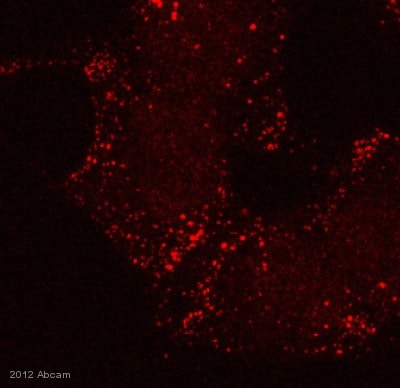
Rat astrocytes stained with fluorescently labeled Caveolin-1 antibody.
Primary antibody is ab2910 at a dilution of 1/500 and the secondary antibody is Texas red labeled anti-rabbit IgG at a dilution of 1/1000.
This image was kindly supplied as part of the review submitted by Donghui Zhu.
Immunocytochemistry/ Immunofluorescence - Anti-Caveolin-1 antibody - Caveolae Marker (ab2910)This image is courtesy of an anonymous Abreview
ab2910 staining Caveolin-1 - Caveolae Marker in HeLa (Human epithelial cell line from cervix adenocarcinoma) cells by ICC/IF (Immunocytochemistry/immunofluorescence).
Cells were fixed with paraformaldehyde. Samples were incubated with primary antibody (1/200 in PBS + 0.05% Saponin) for 1 hour at 37°C. A Cy3®-conjugated Donkey anti-rabbit polyclonal (1/500) was used as the secondary antibody.


