Anti-Ubiquitin抗体
参阅全部 Ubiquitin 一抗
兔多克隆抗体to Ubiquitin
Rabbit
From Mar 2024, QC testing of replenishment batches of this polyclonal changed. All tested and expected application and reactive species combinations are still covered by our Abcam product promise. However, we no longer test all applications. For more information on a specific batch, please contact our Scientific Support who will be happy to help. You may also be interested in our alternative recombinant antibody, ab134953.
适用于: IHC-P, ICC/IF, IHC-FrFlmore details
与反应: Rat, Human
预测可用于: Mouse, Horse, Cow, Monkey, African green monkey![]()
Recombinant full length protein corresponding to Human Ubiquitin.
This antibody stains the periphery of senile plaques and of neurofibrillary tangles in Alzheimer's disease brain, the Lewy bodies in Parkinson's disease brain, and the Mallory bodies in alcoholic liver disease.
This product is FOR RESEARCH USE ONLY. For commercial use, please contact partnerships@abcam.com.
The Life Science industry has been in the grips of a reproducibility crisis for a number of years. Abcam is leading the way in addressing this with our range of recombinant monoclonal antibodies and knockout edited cell lines for gold-standard validation. Please check that this product meets your needs before purchasing.
If you have any questions, special requirements or concerns, please send us an inquiry and/or contact our Support team ahead of purchase. Recommended alternatives for this product can be found below, along with publications, customer reviews and Q&As
Liquid
Shipped at 4°C. Store at +4°C short term (1-2 weeks). Upon delivery aliquot. Store at -20°C. Avoid freeze / thaw cycle.
pH: 7.60
Preservative: 0.1% Sodium azide
Constituents: PBS, 1% BSA
Immunogen affinity purified
多克隆
IgG
Abpromise™承诺保证使用ab7780于以下的经测试应用
“应用说明”部分 下显示的仅为推荐的起始稀释度;实际最佳的稀释度/浓度应由使用者检定。
| 应用 | Ab评论 | 说明 |
|---|---|---|
| IHC-P | (3) | 1/50. Perform heat mediated antigen retrieval before commencing with IHC staining protocol. Boil tissue sections in 10mM citrate buffer, pH 6.0 for 10 min followed by cooling at RT for 20 min. |
| ICC/IF | (7) | Use at an assay dependent concentration. |
| IHC-FrFl | 1/1000. |
Entrez Gene: 7314 Human
Entrez Gene: 22187 Mouse
SwissProt: P0CG47 Human
SwissProt: P0CG48 Human
SwissProt: P62979 Human
SwissProt: P62987 Human
SwissProt: P62988 Human
SwissProt: P0CG49 Mouse
SwissProt: P62991 Mouse
Unigene: 356190 Human
Unigene: 520348 Human
Unigene: 282093 Mouse
Unigene: 371592 Mouse
Unigene: 110618 Rat
Unigene: 1253 Rat
Unigene: 3761 Rat
Epididymis secretory protein Li 50 antibody
FLJ25987 antibody
HEL S 50 antibody
MGC8385 antibody
Polyubiquitin B antibody
RPS 27A antibody
RPS27A antibody
UBA 52 antibody
UBA 80 antibody
UBA52 antibody
UBA80 antibody
UBB antibody
UBB_HUMAN antibody
UBC antibody
UBCEP 1 antibody
UBCEP 2 antibody
UBCEP1 antibody
UBCEP2 antibody
Ubiquitin antibody
Ubiquitin B antibody

Immunocytochemistry/ Immunofluorescence - Anti-Ubiquitin antibody (ab7780)Image courtesy of Armen Petrosyan by Abreview.
ab7780 staining Ubiquitin in Panc-1 cells by Immunocytochemistry/ Immunofluorescence.Cells were fixed in formaldehyde, blocked with 1% serum for 1 hour at 22°C and then incubated with ab7780 at a 1/2000 dilution for 1 hour at 22°C. The secondary was used at a 1/200 dilution.Green - Golgi residental proteins C2GnT-M.Red -Ubiquitin.Blue - nucleus staining by DAPI.

Immunocytochemistry/ Immunofluorescence - Anti-Ubiquitin antibody (ab7780)Zhao A et al., PLoS One., 2013: 8(2): e56203. Fig 4.; doi: 10.1371/journal.pone.0056203 Reproduced under the Creative Commons license http://creativecommons.org/licenses/by/4.0/
ab7780 staining ubiquitin in cells transfected with FGAMS-EGFP by ICC/IF (immunocytochemistry/immunofluorescence). Cells were fixed with 3.7% methanol-free formaldehyde at 37°C for 15-20 minutes, blocked with 5% goat serum in PBS-T buffer for 30-60 minutes at room temperature. An Alexa Fluor® 594-conjugated Goat anti-rabbit polyclonal was used as the secondary antibody

Immunocytochemistry/ Immunofluorescence - Anti-Ubiquitin antibody (ab7780)
ICC/IF image of ab7780 stained MCF7 cells. The cells were 4% PFA fixed (10 min) and then incubated in 1%BSA / 10% normal goat serum (ab7481) / 0.3M glycine in 0.1% PBS-Tween for 1h to permeabilise the cells and block non-specific protein-protein interactions. The cells were then incubated with the antibody (ab7780, 1æg/ml) overnight at +4øC. The secondary antibody (green)ÿwas Alexa Fluor© 488 goat anti-mouse IgG (H+L) ab150113) used at a 1/1000 dilution for 1h. Alexa Fluor© 594 WGA was used to label plasma membranes (red) at a 1/200 dilution for 1h. DAPI was used to stain the cell nuclei (blue) at a concentration of 1.43æM.

Immunohistochemistry (Formalin/PFA-fixed paraffin-embedded sections) - Anti-Ubiquitin antibody (ab7780)
ab7780 (1/100) staining Ubiquitin in paraffin-embedded brain sections of a patient with Alzheimer's disease.

Immunohistochemistry (Formalin/PFA-fixed paraffin-embedded sections) - Anti-Ubiquitin antibody (ab7780)This image is courtesy of an anonymous Abreview
ab7780 staining ubiquitin in Mouse kidney tissue sections by Immunohistochemistry (IHC-P - paraformaldehyde-fixed, paraffin-embedded sections). Tissue was fixed with formaldehyde and blocked with 1% BSA for 1 hour at 25°C; antigen retrieval was by heat mediation in a citrate buffer. Samples were incubated with primary antibody (1/90) for 16 hours at 4°C. A HRP-conjugated Goat anti-rabbit polyclonal (1/1000) was used as the secondary antibody.
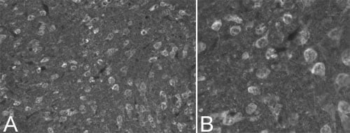
Immunohistochemistry - Free Floating - Anti-Ubiquitin antibody (ab7780)
ab7780 at a dilution of 1/1000, staining Ubiquitin in neurons (Alexa 488 secondary at 1/2000) on 30µm coronal rat brain (ab29475) tissue sections in free floating IHC (see protocol link for detailed description). Image shows neuronal staining observed with [A] 20x objective and [B] 40x objective.
NB: No labeling observed following omission of primary antibody.
Sections were viewed using an Axioplan 2 Imaging microscope (Imaging Associates) fitted with 10x, 20x and 40x Plan-Neofluorobjectives (Zeiss, Germany) and images were taken using a AxioCam Hrm digital camera (Zeiss, Germany) and AxioVision software (Imaging Associates).


