Anti-Histone H3 (di methyl K4)抗体- ChIP Grade
参阅全部 Histone H3 一抗
兔多克隆抗体to Histone H3 (di methyl K4) - ChIP Grade
Rabbit
From Jan 2024, QC testing of replenishment batches of this polyclonal changed. All tested and expected application and reactive species combinations are still covered by our Abcam product promise. However, we no longer test all applications. For more information on a specific batch, please contact our Scientific Support who will be happy to help. You may also be interested in our alternative recombinant antibody, ab176878.
适用于: ChIP, WB, PepArr, ICC/IFmore details
与反应: Mouse, Rat, Cow, Human
预测可用于: Pig, Saccharomyces cerevisiae, Tetrahymena, Drosophila melanogaster, Schizosaccharomyces pombe, Mammals, Plasmodium falciparum, Common marmoset, Candida albicans![]()
Synthetic peptide. This information is proprietary to Abcam and/or its suppliers.
(Peptide available as ab7768)
ChIP: Chromatin prepared from HeLa cells. WB: HeLa, NIH/3T3 and PC12 nuclear lysate (triton enriched). Calf Thymus Histone Preparation Nuclear Lysate. ICC/IF: HeLa cells.
For detection of Histone H3 specifically methylated at position Lys 4. This antibody was used in a screen by Dover et al, (2002) to isolate yeast mutants that are unable to methylate Lysine 4. In immunofluorescence, this antibody detects foci in the nucleus that are non-colocalising with condensed chromatin. The perinuclear and perinucleolar heterochromatin are not stained with this antibody.
Learn about ChIP assay kits, other ChIP antibodies, protocols and more in the ChIP assay guide.
The Life Science industry has been in the grips of a reproducibility crisis for a number of years. Abcam is leading the way in addressing this with our range of recombinant monoclonal antibodies and knockout edited cell lines for gold-standard validation. Please check that this product meets your needs before purchasing.
If you have any questions, special requirements or concerns, please send us an inquiry and/or contact our Support team ahead of purchase. Recommended alternatives for this product can be found below, along with publications, customer reviews and Q&As
Liquid
Shipped at 4°C. Store at +4°C short term (1-2 weeks). Upon delivery aliquot. Store at -20°C or -80°C. Avoid freeze / thaw cycle.
pH: 7.40
Preservative: 0.02% Sodium azide
Constituents: 98.98% PBS, 1% BSA
Batches of this product that have a concentration < 1mg/ml may have BSA added as a stabilising agent. If you would like information about the formulation of a specific lot, please contact our scientific support team who will be happy to help.
Immunogen affinity purified
For detection of Histone H3 specifically methylated at position Lys 4. This antibody was used in a screen by Dover et al, (2002) to isolate yeast mutants that are unable to methylate Lysine 4. In immunofluorescence, this antibody detects foci in the nucleus that are non-colocalising with condensed chromatin. The perinuclear and perinucleolar heterochromatin are not stained with this antibody.
多克隆
IgG
Abpromise™承诺保证使用ab7766于以下的经测试应用
“应用说明”部分 下显示的仅为推荐的起始稀释度;实际最佳的稀释度/浓度应由使用者检定。
| 应用 | Ab评论 | 说明 |
|---|---|---|
| ChIP | (3) | Use 2 µg for 25 µg of chromatin. Use ALDOA ChIP primer pair ab269260 as positive control. |
| WB | (11) | Use a concentration of 1 µg/ml. Detects a band of approximately 17 kDa (predicted molecular weight: 15 kDa). |
| PepArr | Use a concentration of 0.2 - 2 µg/ml. | |
| ICC/IF | (6) | Use a concentration of 5 µg/ml. |
Entrez Gene: 8350 Human
Entrez Gene: 8351 Human
Entrez Gene: 8352 Human
Entrez Gene: 8353 Human
Entrez Gene: 8354 Human
Entrez Gene: 8355 Human
Entrez Gene: 8356 Human
Entrez Gene: 8357 Human
Entrez Gene: 8358 Human
Entrez Gene: 8968 Human
Entrez Gene: 319152 Mouse
Entrez Gene: 319153 Mouse
Entrez Gene: 360198 Mouse
Entrez Gene: 97908 Mouse
Omim: 602810 Human
SwissProt: P68431 Human
SwissProt: P68433 Mouse
Unigene: 132854 Human
Unigene: 247813 Human
Unigene: 247814 Human
Unigene: 248176 Human
Unigene: 443021 Human
Unigene: 484990 Human
Unigene: 532144 Human
Unigene: 533292 Human
Unigene: 546315 Human
Unigene: 586261 Human
Unigene: 591778 Human
Unigene: 221301 Mouse
Unigene: 261657 Mouse
Unigene: 377874 Mouse
Unigene: 390558 Mouse
Unigene: 397328 Mouse
Unigene: 138090 Rat
H3 histone family member E pseudogene antibody
H3 histone family, member A antibody
H3/A antibody
H31_HUMAN antibody
H3F3 antibody
H3FA antibody
Hist1h3a antibody
HIST1H3B antibody
HIST1H3C antibody
HIST1H3D antibody
HIST1H3E antibody
HIST1H3F antibody
HIST1H3G antibody
HIST1H3H antibody
HIST1H3I antibody
HIST1H3J antibody
HIST3H3 antibody
histone 1, H3a antibody
Histone cluster 1, H3a antibody
Histone H3 3 pseudogene antibody
Histone H3.1 antibody
Histone H3/a antibody
Histone H3/b antibody
Histone H3/c antibody
Histone H3/d antibody
Histone H3/f antibody
Histone H3/h antibody
Histone H3/i antibody
Histone H3/j antibody
Histone H3/k antibody
Histone H3/l antibody

ChIP - Anti-Histone H3 (di methyl K4) antibody - ChIP Grade (ab7766)
Chromatin was prepared from HeLa cells according to the Abcam X-ChIP protocol. Cells were fixed with formaldehyde for 10 minutes. The ChIP was performed with 25µg of chromatin, 2µg of ab7766 (blue), and 20µl of Protein A/G sepharose beads. No antibody was added to the beads control (yellow). The immunoprecipitated DNA was quantified by real time PCR (Taqman approach for active and inactive loci, Sybr green approach for heterochromatic loci). Primers and probes are located in the first kb of the transcribed region.

Peptide Array - Anti-Histone H3 (di methyl K4) antibody - ChIP Grade (ab7766)
All batches of ab7766 are tested in Peptide Array against peptides to different Histone H3 modifications. Six dilutions of each peptide are printed on to the Peptide Array in triplicate and results are averaged before being plotted on to a graph. Results show strong binding to Histone H3 - di methyl K4 peptide (ab7768), indicating that this antibody specifically recognises the Histone H3 - di methyl K4 modification.
ab1340 - Histone H3 - mono methyl K4
ab1342 - Histone H3 - tri methyl K4
ab1771 - Histone H3 - mono methyl K9
ab1772 - Histone H3 - di methyl K9
ab1773 - Histone H3 - tri methyl K9
ab1780 - Histone H3 - mono methyl K27
ab1781 - Histone H3 - di methyl K27
ab1782 - Histone H3 - tri methyl K27
ab7228 - Histone H3 - unmodified
ab7768 - Histone H3 - di methyl K4

Immunocytochemistry/ Immunofluorescence - Anti-Histone H3 (di methyl K4) antibody - ChIP Grade (ab7766)
ab7766 staining Histone H3 (di methyl K4) in HeLa cells. The cells were fixed with 4% paraformaldehyde (10 min), permeabilized with 0.1% PBS-Triton X-100 for 5 minutes and then blocked with 1% BSA/10% normal goat serum/0.3M glycine in 0.1% PBS-Tween for 1h. The cells were then incubated overnight at 4°C with ab7766 at 5 µg/ml and ab7291, Mouse monoclonal [DM1A] to alpha Tubulin - Loading Control. Cells were then incubated with ab150081, Goat polyclonal Secondary Antibody to Rabbit IgG - H&L (Alexa Fluor® 488), pre-adsorbed at 1/1000 dilution (shown in green) and ab150120, Goat polyclonal Secondary Antibody to Mouse IgG - H&L (Alexa Fluor® 594), pre-adsorbed at 1/1000 dilution (shown in pseudocolour red). Nuclear DNA was labelled with DAPI (shown in blue).
Also suitable in cells fixed with 100% methanol (5 min).
Image was acquired with a high-content analyser (Operetta CLS, Perkin Elmer) and a maximum intensity projection of confocal sections is shown.
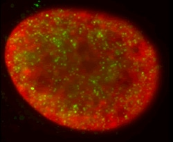
Immunocytochemistry/ Immunofluorescence - Anti-Histone H3 (di methyl K4) antibody - ChIP Grade (ab7766)This image is courtesy of Kirk McManus in the lab of Michael Hendzel, Univeristy of Alberta
Dimethylated lysine 4 (green) is found in several hundred small nuclear foci that do not colocalize with condensed regions of chromatin (DAPI stained, red). The perinuclear and perinucleolar heterochromatin do not stain with this antibody.

Western blot - Anti-Histone H3 (di methyl K4) antibody - ChIP Grade (ab7766)
Anti-Histone H3 (di methyl K4) antibody - ChIP Grade (ab7766) at 1 µg/ml + Calf Thymus Histone Preparation Nuclear Lysate (ab121) at 0.5 µg
Secondary
Goat Anti-Rabbit IgG H&L (HRP) preadsorbed (ab97080) at 1/5000 dilution
Developed using the ECL technique.
Performed under reducing conditions.
Predicted band size: 15 kDa
Observed band size: 17 kDawhy is the actual band size different from the predicted?
Additional bands at: 14 kDa. We are unsure as to the identity of these extra bands.
Exposure time: 4 minutes

Immunocytochemistry/ Immunofluorescence - Anti-Histone H3 (di methyl K4) antibody - ChIP Grade (ab7766)This image is courtesy of an Abreview submitted by Dr Alexander Rapp
ab7766 at 1/200 staining human U2OS (osteosarcoma) cells by ICC/IF. The cells were paraformaldehyde fixed and then stained with the antibody for 1 hour. A Cy2 ® conjugated donkey anti-rabbit antibody was used as the secondary (green). The image shows uniformal staining of the whole nucleus, with several specles found. The insert shows H3-di methyl K4 of tw2 cells only. DAPI nuclear staining is shown in blue.

Western blot - Anti-Histone H3 (di methyl K4) antibody - ChIP Grade (ab7766)
All lanes : Anti-Histone H3 (di methyl K4) antibody - ChIP Grade (ab7766) at 1 µg/ml
Lane 1 : HeLa nuclear lysate (triton enriched)
Lane 2 : NIH 3T3 nuclear lysate (triton enriched)
Lane 3 : PC12 nuclear lysate (triton enriched
Lysates/proteins at 20 µg per lane.
Secondary
All lanes : Goat polyclonal to Rabbit IgG - H&L - Pre-Adsorbed (HRP) at 1/50000 dilution
Predicted band size: 15 kDa
Observed band size: 17 kDawhy is the actual band size different from the predicted?
Blocking buffer: 2% BSA
Gel type: MES
Exposure Time: 1 minute

Western blot - Anti-Histone H3 (di methyl K4) antibody - ChIP Grade (ab7766)
MCF7 cells were incubated at 37°C for 24h with vehicle control (0 µM) and different concentrations of tranylcypromine hydrochloride (ab120606). Increased expression of Histone 3 K4 di-methyl (ab7766) in MCF7 cells correlates with an increase in tranylcypromine hydrochloride concentration, as described in literature.
Nuclear extracts were prepared with RIPA buffer (containing protease inhibitors and sodium orthovanadate), 10 µg of each were loaded on the gel and the WB was run under reducing conditions. After transfer the membrane was blocked for an hour using 5% BSA before being incubated with ab7766 at 1 µg /ml and ab1791 at 1 µg /ml overnight at 4°C. Antibody binding was detected using an anti-rabbit HRP secondary antibody (ab97051) at 1/10000 dilution and visualized using ECL development solution.


