Anti-Bim抗体
参阅全部 Bim 一抗
兔多克隆抗体to Bim
Rabbit
适用于: IHC-P, WB, ICC/IFmore details
与反应: Rat, Human
Peptide corresponding to amino acids 22 to 40 of human origin. The sequence is identical to that of mouse and differs from that of rat by one amino acid.
WB: Human colon and thymus tissue lysates, Rat Myeloma Cell lysate ICC/IF: K562 cells
The Life Science industry has been in the grips of a reproducibility crisis for a number of years. Abcam is leading the way in addressing this with our range of recombinant monoclonal antibodies and knockout edited cell lines for gold-standard validation. Please check that this product meets your needs before purchasing.
If you have any questions, special requirements or concerns, please send us an inquiry and/or contact our Support team ahead of purchase. Recommended alternatives for this product can be found below, along with publications, customer reviews and Q&As
Liquid
Shipped at 4°C. Upon delivery aliquot and store at -20°C or -80°C. Avoid repeated freeze / thaw cycles.
pH: 7.2
Preservative: 0.02% Sodium azide
浓度
100 µg 浓度为 1 mg/ml
Affinity purified
多克隆
IgG
Abpromise™承诺保证使用ab7888于以下的经测试应用
“应用说明”部分 下显示的仅为推荐的起始稀释度;实际最佳的稀释度/浓度应由使用者检定。
| 应用 | Ab评论 | 说明 |
|---|---|---|
| IHC-P | Use a concentration of 20 µg/ml. | |
| WB | Use a concentration of 1 µg/ml. Predicted molecular weight: 23 kDa. | |
| ICC/IF | Use at an assay dependent concentration. |
Entrez Gene: 10018 Human
Omim: 603827 Human
SwissProt: O43521 Human
Unigene: 469658 Human
Unigene: 82709 Rat
Bcl 2 interacting protein Bim antibody
Bcl 2 related ovarian death agonist antibody
Bcl-2-like protein 11 antibody
BCL2 interacting mediator of cell death antibody
BCL2 like 11 (apoptosis facilitator) antibody
BCL2 like protein 11 antibody
Bcl2-interacting mediator of cell death antibody
Bcl2-L-11 antibody
Bcl2l11 antibody
BIM alpha6 antibody
BIM antibody
BIM beta6 antibody
BIM beta7 antibody
BimEL antibody
BimL antibody
BOD antibody
BCL2 like 11 antibody
B2L11_HUMAN antibody
BAM antibody

Western blot - Anti-Bim antibody (ab7888)
All lanes : Anti-Bim antibody (ab7888) at 0.5 µg/ml
Lane 1 : Human thymus tissue lysate
Lane 2 : Human colon tissue lysate
Lysates/proteins at 15 µg per lane.
Secondary
All lanes : Goat anti-rabbit IgG HRP conjugate at 1/10000 dilution
Predicted band size: 23 kDa

Immunohistochemistry (Formalin/PFA-fixed paraffin-embedded sections) - Anti-Bim antibody (ab7888)
ab7888 at 20µg/ml staining Bim in human cancer cells by IHC-P
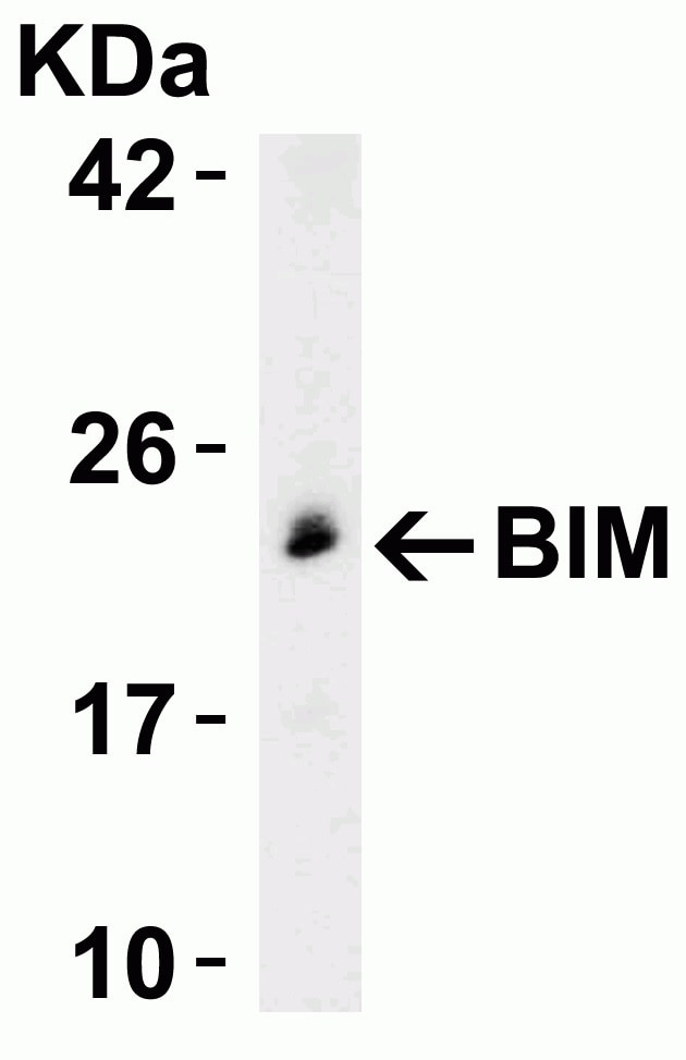
Western blot - Anti-Bim antibody (ab7888)
Anti-Bim antibody (ab7888) at 0.5 µg/ml + Rat Myeloma Cell lysate at 15 µg
Secondary
Goat anti-rabbit IgG HRP conjugate at 1/10000 dilution
Predicted band size: 23 kDa

Immunocytochemistry/ Immunofluorescence - Anti-Bim antibody (ab7888)
Immunocytochemistry/ Immunofluorescence analysis of 4% paraformaldehyde fixed human K562 cells labeling Bim with ab7888 at 20 μg/mL. Goat anti-rabbit IgG secondary antibody at 1/500 dilution was used as the secondry antibody.

Immunocytochemistry/ Immunofluorescence - Anti-Bim antibody (ab7888)
ICC/IF image of ab7888 stained HepG2 cells. The cells were 4% formaldehyde (10 min) and then incubated in 1%BSA / 10% normal goat serum / 0.3M glycine in 0.1% PBS-Tween for 1h to permeabilise the cells and block non-specific protein-protein interactions. The cells were then incubated with the antibody (ab7888, 5µg/ml) overnight at +4°C. The secondary antibody (green) was ab96899 Dylight 488 goat anti-rabbit IgG (H+L) used at a 1/250 dilution for 1h. Alexa Fluor® 594 WGA was used to label plasma membranes (red) at a 1/200 dilution for 1h. DAPI was used to stain the cell nuclei (blue) at a concentration of 1.43µM.


