Anti-FAK (phospho Y861)抗体
参阅全部 FAK 一抗
兔多克隆抗体to FAK (phospho Y861)
Rabbit
适用于: WB, ICCmore details
与反应: Chicken, Human
预测可用于: Rat, Xenopus laevis![]()
Synthetic peptide corresponding to Human FAK (phospho Y861). The sequence is conserved in mouse, rat, chicken and frog.
Focal Adhesion Kinase is a 125 kDa non-receptor protein tyrosine kinase that is a substrate for Src and a key element in growth factor and integrin signalling. Focal Adhesion Kinase plays a central role in cell spreading, differentiation, migration, cell death and acceleration of the G1 to S phase transition of the cell cycle. Tyr861 of Focal Adhesion Kinase is a major Src phosphorylation site that allows Focal Adhesion Kinase to bind to integrins and is also involved in cancer.
The Life Science industry has been in the grips of a reproducibility crisis for a number of years. Abcam is leading the way in addressing this with our range of recombinant monoclonal antibodies and knockout edited cell lines for gold-standard validation. Please check that this product meets your needs before purchasing.
If you have any questions, special requirements or concerns, please send us an inquiry and/or contact our Support team ahead of purchase. Recommended alternatives for this product can be found below, along with publications, customer reviews and Q&As
Liquid
Shipped at 4°C. Upon delivery aliquot and store at -20°C or -80°C. Avoid repeated freeze / thaw cycles.
pH: 7.3
Preservative: 0.05% Sodium azide
Constituents: PBS, 50% Glycerol (glycerin, glycerine), 0.1% BSA
BSA is IgG and protease free
浓度
批次浓度范围 50 µl 浓度为 0.25 - 0.4 mg/ml
Immunogen affinity purified
Purified from rabbit serum by sequential epitope-specific chromatography. The antibody has been negatively preadsorbed using (i) a non-phosphopeptide corresponding to the site of phosphorylation to remove antibody that is reactive with non-phosphorylated Focal Adhesion Kinase protein, and (ii) a generic tyrosine phosphorylated peptide to remove antibody that is reactive with phosphotyrosine (irrespective of the sequence). The final product is generated by affinity chromatography using a Focal Adhesion Kinase-derived peptide that is phosphorylated at tyrosine 861.
Focal Adhesion Kinase is a 125 kDa non-receptor protein tyrosine kinase that is a substrate for Src and a key element in growth factor and integrin signalling. Focal Adhesion Kinase plays a central role in cell spreading, differentiation, migration, cell death and acceleration of the G1 to S phase transition of the cell cycle. Tyr861 of Focal Adhesion Kinase is a major Src phosphorylation site that allows Focal Adhesion Kinase to bind to integrins and is also involved in cancer.
多克隆
IgG
Abpromise™承诺保证使用ab4804于以下的经测试应用
“应用说明”部分 下显示的仅为推荐的起始稀释度;实际最佳的稀释度/浓度应由使用者检定。
| 应用 | Ab评论 | 说明 |
|---|---|---|
| WB | (2) | 1/1000. Predicted molecular weight: 119 kDa. |
| ICC | 1/250. |
Entrez Gene: 396416 Chicken
Entrez Gene: 5747 Human
Entrez Gene: 399286 Xenopus laevis
Omim: 600758 Human
SwissProt: Q00944 Chicken
SwissProt: Q05397 Human
SwissProt: Q91738 Xenopus laevis
Unigene: 395482 Human
Unigene: 2809 Rat
Unigene: 6819 Xenopus laevis
FADK 1 antibody
FADK antibody
FAK related non kinase polypeptide antibody
FAK1 antibody
FAK1_HUMAN antibody
Focal adhesion kinase 1 antibody
Focal adhesion Kinase antibody
Focal adhesion kinase isoform FAK Del33 antibody
Focal adhesion kinase related nonkinase antibody
FRNK antibody
p125FAK antibody
pp125FAK antibody
PPP1R71 antibody
Protein phosphatase 1 regulatory subunit 71 antibody
Protein tyrosine kinase 2 antibody
Protein-tyrosine kinase 2 antibody
Ptk2 antibody
PTK2 protein tyrosine kinase 2 antibody

Immunocytochemistry - Anti-FAK (phospho Y861) antibody (ab4804)
Immunofluorescence analysis of FAK [pY861] was done on 70% confluent log phase A-549 cells. The cells were fixed with 4% paraformaldehyde for 15 minutes, permeabilized with 0.25% Triton™ X-100 for 10 minutes, and blocked with 5% BSA for 1 hour at room temperature. The cells were labelled with ab4804 at 1:250 dilution in 1% BSA and incubated for 3 hours at room temperature and then labelled with a Goat anti-Rabbit IgG (H+L) Superclonal™ Secondary Antibody, Alexa Fluor® 488 conjugate at a dilution of 1:2000 for 45 minutes at room temperature (Panel a: green). Nuclei (Panel b: blue) were stained with DAPI. F-actin (Panel c: red) was stained with Rhodamine Phalloidin at a 1:300 dilution. Panel d is a merged image showing localization of target protein at focal adhesions. Panel e is a no primary antibody control. The images were captured at 60X magnification.

Western blot - Anti-FAK (phospho Y861) antibody (ab4804)
Peptide Competition: Cell extracts prepared from chick embryo fibroblasts expressing FAK and plated on fibronectin were resolved by SDS-PAGE on a 10% Tris-glycine gel. The proteins then were transferred to nitrocellulose and incubated with 0.50 µg/mL ab4804 antibody, following prior incubation with: (1) the phosphopeptide immunogen, (2) a generic phosphotyrosine containing peptide, (3) the non-phosphorylated peptide corresponding to the phosphopeptide, and (4) no peptide. After washing, membranes were incubated with goat F(ab’)2 anti-rabbit IgG alkaline phosphatase and bands were detected using the Tropix WesternStar detection method. The data show that only the phosphopeptide corresponding to this site blocks the antibody signal, demonstrating the specificity of the ab4804 antibody for this phosphorylated residue. Peptide Competition: Cell extracts prepared from chick embryo fibroblasts expressing FAK and plated on fibronectin were resolved by SDS-PAGE on a 10% T
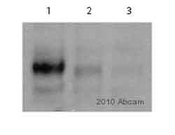
Western blot - Anti-FAK (phospho Y861) antibody (ab4804)Image courtesy of an anonymous Abreview.
All lanes : Anti-FAK (phospho Y861) antibody (ab4804) at 1/1000 dilution
Lane 1 : Whole cell lysate prepared from human MDA-MB-231 breast cancer cells, un-treated
Lane 2 : Whole cell lysate prepared from human MDA-MB-231 breast cancer cells, treated for 1hr with 2.5uM AZD0530 src inhibitor
Lane 3 : Whole cell lysate prepared from human MDA-MB-231 breast cancer cells, treated for 1hr with 5uM AZD0530 src inhibitor
Lysates/proteins at 25 µg per lane.
Secondary
All lanes : Goat anti-rabbit HRP conjugated at 1/5000 dilution
Developed using the ECL technique.
Performed under reducing conditions.
Predicted band size: 119 kDa
Observed band size: 125 kDawhy is the actual band size different from the predicted?
Exposure time: 5 minutes
Primary antibody incubated for 16 hours at 4°C.
Blocking step was performed using 5% milk for 1 hour at 25°C.


