Anti-CYP2C11抗体
兔多克隆抗体to CYP2C11
Rabbit
适用于: IHC-P, ICC/IFmore details
与反应: Rat, Human
This product was produced with the following immunogens:
Synthetic peptide corresponding to Rat CYP2C11 aa 1-100.
Synthetic peptide corresponding to Rat CYP2C11 aa 450-550.
The Life Science industry has been in the grips of a reproducibility crisis for a number of years. Abcam is leading the way in addressing this with our range of recombinant monoclonal antibodies and knockout edited cell lines for gold-standard validation. Please check that this product meets your needs before purchasing.
If you have any questions, special requirements or concerns, please send us an inquiry and/or contact our Support team ahead of purchase. Recommended alternatives for this product can be found below, along with publications, customer reviews and Q&As
Liquid
Shipped at 4°C. Store at +4°C short term (1-2 weeks). Upon delivery aliquot. Store at -20°C or -80°C. Avoid freeze / thaw cycle.
Preservative: 0.05% Sodium azide
Constituent: 99% PBS
Whole antiserum
The Cytochrome P450 (P450) family of enzymes is one of three enzyme systems which metabolize the fatty acid arachadonic acid (AA) to regulators of vascular tone. P450 enzymes are monooxygenase enzymes which require several co-factors such as NADPH and P450 reductase. There are over 200 cDNA’s which encode P450 protein. Epoxygenases are those P450 proteins which metabolize AA to epoxyeicosatrienoic acid (EETs) and omega-hydroxylases are those P450 proteins which produce 19- and 20-hydroxyeicosatetraenoic acids (19- and 20-HETE). EET’s, which exhibit vasodilation activity, are formed when an epoxide group is inserted between the unsaturated carbons of AA in positions 5,6; 8,9; 11,12; 14,15. EET’s are produced in cerebral cortical tissue, coronary arteries and vascular endothelium. EET’s are converted from AA by the 2C11 family of P450’s whose expression is induced by testosterone and is therefore not generally found in females.
多克隆
IgG
Abpromise™承诺保证使用ab3571于以下的经测试应用
“应用说明”部分 下显示的仅为推荐的起始稀释度;实际最佳的稀释度/浓度应由使用者检定。
| 应用 | Ab评论 | 说明 |
|---|---|---|
| IHC-P | 1/100 - 1/500. Perform heat mediated antigen retrieval with citrate buffer pH 6 before commencing with IHC staining protocol. | |
| ICC/IF | 1/20 - 1/200. |
CYP2CII antibody
CYPIIC11 antibody
Cytochrome P-450(M-1) antibody
Cytochrome P450 2C11 antibody
Cytochrome P450-UT-2 antibody
Cytochrome P450-UT-A antibody
Cytochrome P450H antibody
P450 UT A antibody
P450H antibody
UT2 antibody
CP2CB_RAT antibody
Cyp2c antibody
Cyp2c11 antibody

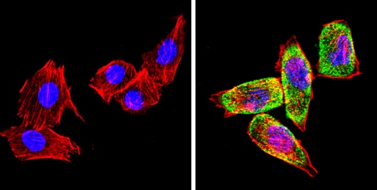
Immunocytochemistry/ Immunofluorescence - Anti-CYP2C11 antibody (ab3571)
ab3571 staining CYP2C11 (green) in HeLa (Human epithelial adenocarcinoma cell line) cells (right), compared to control (left). Formalin-fixed cells were permeabilized with 0.1% Triton X-100 in TBS for 5-10 minutes and blocked with 3% BSA-PBS for 30 minutes at room temperature. Cells were incubated with primary antibody (1:100 in 3% BSA-PBS) overnight at 4 ºC. A DyLight-conjugated anti-rabbit was used as the secondary antibody. Red (phalloidin) - F-actin, Blue - nuclei. Images were taken at a magnification of 60x.

Immunohistochemistry (Formalin/PFA-fixed paraffin-embedded sections) - Anti-CYP2C11 antibody (ab3571)
ab3571 staining CYP2C11 in the cytoplasm of rat liver tissue (right) compared with a negative control in the absence of primary antibody (left) by Immunohistochemistry (Formalin/PFA-fixed paraffin-embedded sections). To expose target proteins, antigen retrieval method was performed using 10mM sodium citrate (pH 6.0) microwaved for 8-15 min. Tissues were then blocked in 3% H2O2-methanol for 15 min at room temperature. Sections were incubated with primary antibody (1:200 in 3% BSA-PBS) overnight at 4°C. A HRP-conjugated anti-rabbit was used as the secondary antibody, followed by colorimetric detection using a DAB kit. Tissues were counterstained with hematoxylin and dehydrated with ethanol and xylene to prep for mounting.

Immunocytochemistry/ Immunofluorescence - Anti-CYP2C11 antibody (ab3571)
ab3571 staining CYP2C11 (green) in PC-12 (Rat adrenal gland pheochromocytoma cell line) cells (right), compared to control (left). Formalin-fixed cells were permeabilized with 0.1% Triton X-100 in TBS for 5-10 minutes and blocked with 3% BSA-PBS for 30 minutes at room temperature. Cells were incubated with primary antibody (1:100 in 3% BSA-PBS) overnight at 4 ºC. A DyLight-conjugated anti-rabbit was used as the secondary antibody. Red (phalloidin) - F-actin, Blue - nuclei. Images were taken at a magnification of 60x.
Immunocytochemistry/ Immunofluorescence - Anti-CYP2C11 antibody (ab3571)
ab3571 staining CYP2C11 (green) in H-4-II-E (Rat hepatoma cell line) cells (right), compared to control (left). Formalin-fixed cells were permeabilized with 0.1% Triton X-100 in TBS for 5-10 minutes and blocked with 3% BSA-PBS for 30 minutes at room temperature. Cells were incubated with primary antibody (1:100 in 3% BSA-PBS) overnight at 4 ºC. A DyLight-conjugated anti-rabbit was used as the secondary antibody. Red (phalloidin) - F-actin, Blue - nuclei. Images were taken at a magnification of 60x.

Immunohistochemistry (Formalin/PFA-fixed paraffin-embedded sections) - Anti-CYP2C11 antibody (ab3571)
ab3571 staining CYP2C11 in the cytoplasm of rat kidney tissue (right) compared with a negative control in the absence of primary antibody (left) by Immunohistochemistry (Formalin/PFA-fixed paraffin-embedded sections). To expose target proteins, antigen retrieval method was performed using 10mM sodium citrate (pH 6.0) microwaved for 8-15 min. Tissues were then blocked in 3% H2O2-methanol for 15 min at room temperature. Sections were incubated with primary antibody (1:200 in 3% BSA-PBS) overnight at 4°C. A HRP-conjugated anti-rabbit was used as the secondary antibody, followed by colorimetric detection using a DAB kit. Tissues were counterstained with hematoxylin and dehydrated with ethanol and xylene to prep for mounting.

Immunohistochemistry (Formalin/PFA-fixed paraffin-embedded sections) - Anti-CYP2C11 antibody (ab3571)
IHC image of ab3571 staining in human renal carcinoma formalin fixed paraffin embedded tissue section, performed on a Leica BondTM system using the standard protocol F. The section was pre-treated using heat mediated antigen retrieval with sodium citrate buffer (pH6, epitope retrieval solution 1) for 20 mins. The section was then incubated with ab3571, 5µg/ml, for 15 mins at room temperature and detected using an HRP conjugated compact polymer system. DAB was used as the chromogen. The section was then counterstained with haematoxylin and mounted with DPX.
For other IHC staining systems (automated and non-automated) customers should optimize variable parameters such as antigen retrieval conditions, primary antibody concentration and antibody incubation times.


