Anti-mSin3A抗体- ChIP Grade
参阅全部 mSin3A 一抗
兔多克隆抗体to mSin3A - ChIP Grade
Rabbit
适用于: ChIP, ICC/IF, WB, IHC-Pmore details
与反应: Mouse, Human
Synthetic peptide corresponding to Mouse mSin3A aa 1-100.
The Life Science industry has been in the grips of a reproducibility crisis for a number of years. Abcam is leading the way in addressing this with our range of recombinant monoclonal antibodies and knockout edited cell lines for gold-standard validation. Please check that this product meets your needs before purchasing.
If you have any questions, special requirements or concerns, please send us an inquiry and/or contact our Support team ahead of purchase. Recommended alternatives for this product can be found below, along with publications, customer reviews and Q&As
Liquid
Shipped at 4°C. Store at +4°C short term (1-2 weeks). Upon delivery aliquot. Store at -20°C or -80°C. Avoid freeze / thaw cycle.
Preservative: 0.05% Sodium azide
Constituents: 0.1% BSA, PBS
浓度
100 µg 浓度为 1 mg/ml
Immunogen affinity purified
多克隆
IgG
Abpromise™承诺保证使用ab3479于以下的经测试应用
“应用说明”部分 下显示的仅为推荐的起始稀释度;实际最佳的稀释度/浓度应由使用者检定。
| 应用 | Ab评论 | 说明 |
|---|---|---|
| ChIP | (2) | Use at an assay dependent concentration. |
| ICC/IF | (1) | 1/50 - 1/500. |
| WB | (5) | Use a concentration of 0.5 µg/ml. Detects a band of approximately 150 kDa (predicted molecular weight: 145 kDa). |
| IHC-P | (1) | Use a concentration of 2 µg/ml. |
Entrez Gene: 25942 Human
Entrez Gene: 20466 Mouse
Omim: 607776 Human
SwissProt: Q96ST3 Human
SwissProt: Q60520 Mouse
Unigene: 513039 Human
Unigene: 15755 Mouse
Histone deacetylase complex subunit Sin 3a antibody
Histone deacetylase complex subunit Sin3a antibody
KIAA0700 antibody
Kiaa4126 antibody
mKIAA4126 antibody
Paired amphipathic helix protein Sin 3a antibody
Paired amphipathic helix protein Sin3a antibody
Sin 3a antibody
SIN3 homolog A antibody
SIN3 homolog A transcription regulator (yeast) antibody
SIN3 homolog A transcription regulator antibody
SIN3 transcription regulator homolog A antibody
Sin3a antibody
SIN3A protein antibody
SIN3A_HUMAN antibody
Transcriptional co repressor Sin 3A antibody
Transcriptional co repressor Sin3A antibody
Transcriptional corepressor Sin3a antibody
Transcriptional regulator SIN3A antibody
AW553200 antibody
DKFZP434K2235 antibody
FLJ90319 antibody

Western blot - Anti-mSin3A antibody - ChIP Grade (ab3479)
Western blot of mSin3A on K562 cell extract using ab3479.

Immunohistochemistry (Formalin/PFA-fixed paraffin-embedded sections) - Anti-mSin3A antibody - ChIP Grade (ab3479)
Paraffin-embedded human ovary carcinoma tissue (right panel) stained for mSin3A using ab3479 at 1/200 dilution compared to a negative control without primary antibody (left panel) in immunohistochemical analysis, followed by HRP-conjugated secondary antibody. Detection: DAB staining.
Antigen retrieval was performed using 10mM sodium citrate (pH 6.0), microwaved for 8-15 min.

ChIP - Anti-mSin3A antibody - ChIP Grade (ab3479)
ChIP analysis of Sin3A was performed using cross-linked chromatin from 1x10⁶ HCT 116 (human colorectal carcinoma cell line) cells treated with serum for 0, 15, and 30 minutes. IP was performed using a multiplex microplate Matrix ChIP assay with 1.0 µl/100 µl well volume of ab3479. Chromatin aliquots from ~1x10⁵ cells were used per ChIP pull-down. Quantitative PCR data were done in quadruplicate using 1 µl of eluted DNA in 2 µl SYBR real-time PCR reactions containing primers to amplify -15kb upstream of the Egr1 gene or exon-1 or exon-2 of Egr1. Quantitation of immunoprecipitated chromatin is presented as signal relative to the total amount of input chromatin. A schematic representation of the Egr-1 locus is shown above the data where boxes represent exons (black boxes = translated regions, white boxes = untranslated regions), the zigzag line represents an intron, and the straight line represents upstream sequence. Regions amplified by Egr-1 primers are represented by black bars.

Immunocytochemistry/ Immunofluorescence - Anti-mSin3A antibody - ChIP Grade (ab3479)
Immunocytochemical immunoflurescence analysis of NIH-3T3 cells labelling mSin3A using ab3479. Formalin-fixed cells were permeablized with 0.1% Triton X-100 in TBS for 5-10 minutes and blocked with 3% BSA-PBS for 30 minutes at room temperature. Cells were then incubated with ab3479 in 3% BSA-PBS at a dilution of 1:100 overnight in a 4°C high humidity environment. Cells were then washed with PBST and incubated with a green DyLight-conjugated secondary antibody in PBS at room temperature in the dark. Cells were counterstained with DAPI or Hoechst labelling the nuclear DNA blue. The Left Image is a negative control without the prescence of ab3479. Image magnification is 60X.

Immunocytochemistry/ Immunofluorescence - Anti-mSin3A antibody - ChIP Grade (ab3479)
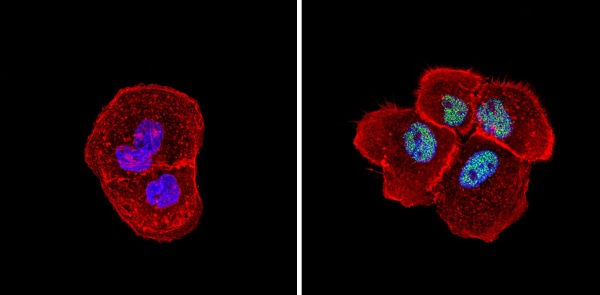
Immunocytochemical immunoflurescence analysis of HeLa cells labelling mSin3A using ab3479. Formalin-fixed cells were permeablized with 0.1% Triton X-100 in TBS for 5-10 minutes and blocked with 3% BSA-PBS for 30 minutes at room temperature. Cells were then incubated with ab3479 in 3% BSA-PBS at a dilution of 1:200 overnight in a 4°C high humidity environment. Cells were then washed with PBST and incubated with a green DyLight-conjugated secondary antibody in PBS at room temperature in the dark. Cells were counterstained blue with DAPI or Hoechst labelling the nuclear DNA and red against actin using an Alexa Fluor® 554 conjugate. The Left Image is a negative control without the prescence of ab3479. Image magnification is 60X.
Immunocytochemistry/ Immunofluorescence - Anti-mSin3A antibody - ChIP Grade (ab3479)
Immunocytochemical immunoflurescence analysis of NIH-3T3 cells labelling mSin3A using ab3479. Formalin-fixed cells were permeablized with 0.1% Triton X-100 in TBS for 5-10 minutes and blocked with 3% BSA-PBS for 30 minutes at room temperature. Cells were then incubated with ab3479 in 3% BSA-PBS at a dilution of 1:100 overnight in a 4°C high humidity environment. Cells were then washed with PBST and incubated with a green DyLight-conjugated secondary antibody in PBS at room temperature in the dark. Cells were counterstained blue with DAPI or Hoechst labelling the nuclear DNA and red against actin using an Alexa Fluor® 554 conjugate. The Left Image is a negative control without the prescence of ab3479. Image magnification is 60X.

Immunohistochemistry (Formalin/PFA-fixed paraffin-embedded sections) - Anti-mSin3A antibody - ChIP Grade (ab3479)
ab3479 (2µg/ml) staining mSin3A in human duodenum using an automated system (DAKO Autostainer Plus). Using this protocol there is strong staining of nuclei of epithelial cells.
Sections were rehydrated and antigen retrieved with the Dako 3 in 1 AR buffers citrate pH6.1/ EDTA pH 9.0 in a DAKO PT link. Slides were peroxidase blocked in 3% H2O2 in methanol for 10 mins. They were then blocked with Dako Protein block for 10 minutes (containing casein 0.25% in PBS) then incubated with primary antibody for 20 min and detected with Dako envision flex amplification kit for 30 minutes. Colorimetric detection was completed with Diaminobenzidine for 5 minutes. Slides were counterstained with Haematoxylin and coverslipped under DePeX. Please note that, for manual staining, optimization of primary antibody concentration and incubation time is recommended. Signal amplification may be required.
抱歉,暂无浏览记录


