Anti-RAGE抗体
参阅全部 RAGE 一抗
兔多克隆抗体to RAGE
Rabbit
By Western blot, this antibody detects two bands in the 45 kDa range representing the RAGE protein pre and post glycosylation in Mouse lung extract. This antibody also detects an ~25 kDa protein that is believed to be proteolytic degradation product. Immunohistochemical staining of RAGE in transgenic Mouse retina results in staining of the retinal pigmented epithelium and photo receptor cell layers.
适用于: IHC-Fr, WB, IHC-Pmore details
与反应: Mouse
Synthetic peptide corresponding to Rat RAGE aa 350-450.
WB: Mouse lung tissue lysate. IHC-P: Mouse lymph node, kidney and heart tissues. IHC-Fr: Transgenic mouse retina.
The Life Science industry has been in the grips of a reproducibility crisis for a number of years. Abcam is leading the way in addressing this with our range of recombinant monoclonal antibodies and knockout edited cell lines for gold-standard validation. Please check that this product meets your needs before purchasing.
If you have any questions, special requirements or concerns, please send us an inquiry and/or contact our Support team ahead of purchase. Recommended alternatives for this product can be found below, along with publications, customer reviews and Q&As
Liquid
Shipped at 4°C. Store at +4°C short term (1-2 weeks). Upon delivery aliquot. Store at -20°C or -80°C. Avoid freeze / thaw cycle.
Preservative: 0.05% Sodium azide
Constituents: 0.1% BSA, 99% PBS
Immunogen affinity purified
多克隆
IgG
Abpromise™承诺保证使用ab3611于以下的经测试应用
“应用说明”部分 下显示的仅为推荐的起始稀释度;实际最佳的稀释度/浓度应由使用者检定。
| 应用 | Ab评论 | 说明 |
|---|---|---|
| IHC-Fr | (1) | Use a concentration of 1 - 2 µg/ml. |
| WB | (6) | Use a concentration of 1 µg/ml. Detects a band of approximately 45 kDa (predicted molecular weight: 42.6 kDa). |
| IHC-P | (1) | 1/10 - 1/100. |
Entrez Gene: 11596 Mouse
SwissProt: Q62151 Mouse
Unigene: 3383 Mouse
MGC2235 antibody
MGC22357 antibody
RAGE_HUMAN antibody
Receptor for advanced glycation end products antibody
Receptor for advanced glycosylation end products antibody
Advanced glycosylation end product-specific receptor antibody
Ager antibody
DAMA 358M23.4 antibody

Western blot - Anti-RAGE antibody (ab3611)
ab3611 at a 2µg/ml concentration staining ~ 45 kDa RAGE in mouse lung lysate by Western blot (ECL).This antibody detects two bands in the 45 kDa range representing the RAGE protein pre and post-glycosylation in mouse lung extract. This antibody also detects an ~25 kDa protein that is believed to be proteolytic degradation product.

Immunohistochemistry (Formalin/PFA-fixed paraffin-embedded sections) - Anti-RAGE antibody (ab3611)
Immunohistochemistry was performed on normal biopsies of deparaffinized Mouse lymph node tissue . To expose target proteins heat induced antigen retrieval was performed using 10mM sodium citrate (pH6.0) buffer microwaved for 8-15 minutes. Following antigen retrieval tissues were blocked in 3% BSA-PBS for 30 minutes at room temperature. Tissues were then probed at a dilution of 1:20 with a rabbit polyclonal antibody recognizing RAGE ab3611 or without primary antibody (negative control) overnight at 4°C in a humidified chamber. Tissues were washed extensively with PBST and endogenous peroxidase activity was quenched with a peroxidase suppressor. Detection was performed using a biotin-conjugated secondary antibody and SA-HRP followed by colorimetric detection using DAB. Tissues were counterstained with hematoxylin and prepped for mounting.
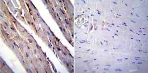
Immunohistochemistry (Formalin/PFA-fixed paraffin-embedded sections) - Anti-RAGE antibody (ab3611)
Immunohistochemistry was performed on normal biopsies of deparaffinized Mouse heart tissue. To expose target proteins heat induced antigen retrieval was performed using 10mM sodium citrate (pH6.0) buffer microwaved for 8-15 minutes. Following antigen retrieval tissues were blocked in 3% BSA-PBS for 30 minutes at room temperature. Tissues were then probed at a dilution of 1:20 with a rabbit polyclonal antibody recognizing RAGE ab3611 or without primary antibody (negative control) overnight at 4°C in a humidified chamber. Tissues were washed extensively with PBST and endogenous peroxidase activity was quenched with a peroxidase suppressor. Detection was performed using a biotin-conjugated secondary antibody and SA-HRP followed by colorimetric detection using DAB. Tissues were counterstained with hematoxylin and prepped for mounting.

Immunohistochemistry (Frozen sections) - Anti-RAGE antibody (ab3611)
Ab3611 used in IHC (frozen) in transgenic mouse retinas.

Immunohistochemistry (Formalin/PFA-fixed paraffin-embedded sections) - Anti-RAGE antibody (ab3611)
Immunohistochemistry was performed on normal biopsies of deparaffinized Mouse kidney tissue. To expose target proteins heat induced antigen retrieval was performed using 10mM sodium citrate (pH6.0) buffer microwaved for 8-15 minutes. Following antigen retrieval tissues were blocked in 3% BSA-PBS for 30 minutes at room temperature. Tissues were then probed at a dilution of 1:20 with a rabbit polyclonal antibody recognizing RAGE ab3611 or without primary antibody (negative control) overnight at 4°C in a humidified chamber. Tissues were washed extensively with PBST and endogenous peroxidase activity was quenched with a peroxidase suppressor. Detection was performed using a biotin-conjugated secondary antibody and SA-HRP followed by colorimetric detection using DAB. Tissues were counterstained with hematoxylin and prepped for mounting.


