| 存储条件 |
|---|
| Product Name | Cytokeratin 7 antibody-Cytoskeleton Marker |
|---|---|
| Antibody Type | Primary Antibodies |
| Antigen Alias | CK 7 antibody CK-7 antibody CK7 antibody Cytokeratin 7 antibody Cytokeratin-7 antibody D15Wsu77e antibody K2C7 antibody K2C7_HUMAN antibody K7 antibody Keratin 7 antibody Keratin 7, type II antibody Keratin type II cytoskeletal 7 antibody Keratin, 55K type II cytoskeletal antibody Keratin, simple epithelial antibody Keratin, simple epithelial type I, K7 antibody Keratin, type II cytoskeletal 7 antibody Keratin-7 antibody Krt2-7 antibody KRT7 antibody MGC11625 antibody MGC129731 antibody MGC3625 antibody Sarcolectin antibody SCL antibody Type II mesothelial keratin K7 antibody Type-II keratin Kb7 antibody |
| Product description | Keratin-7 is a member of the keratin gene family. The type II cytokeratins consist of basic or neutral proteins which are arranged in pairs of heterotypic keratin chains coexpressed during differentiation of simple and stratified epithelial tissues. Keratin-7 is found in simple glandular epithelia, and in transitional epithelium. Epithelial cells of the lung and breast both contain keratin-7, but some other glandular epithelia, such as those of the colon and prostate, do not. Because the keratin-7 antigen is found in both healthy and neoplastic cells, antibodies to CK7 can be used in immunohistochemistry to distinguish ovarian and transitional cell carcinomas from colonic and prostate cancers, respectively. |
| Immunogen | This antibody is produced by immunizing rabbits with a synthetic peptide (KLH-coupled) corresponding to C -terminal cytokeratin 7. |
| Clonality | Polyclonal |
|---|---|
| Isotype | IgG |
| Host Species | Rabbit |
| Tested Applications | WBICCIHCFC |
| WB 1:500: ICC 1:200: IHC 1:200: FC 1:50-1:100: | |
| Species Reactivity | Human |
| Concentration | 1mg/ml |
| Alternative Names | CK 7 antibody CK-7 antibody CK7 antibody Cytokeratin 7 antibody Cytokeratin-7 antibody D15Wsu77e antibody K2C7 antibody K2C7_HUMAN antibody K7 antibody Keratin 7 antibody Keratin 7 type II antibody Keratin type II cytoskeletal 7 antibody Keratin 55K type II cytoskeletal antibody Keratin simple epithelial antibody Keratin simple epithelial type I K7 antibody Keratin type II cytoskeletal 7 antibody Keratin-7 antibody Krt2-7 antibody KRT7 antibody MGC11625 antibody MGC129731 antibody MGC3625 antibody Sarcolectin antibody SCL antibody Type II mesothelial keratin K7 antibody Type-II keratin Kb7 antibody |
|---|---|
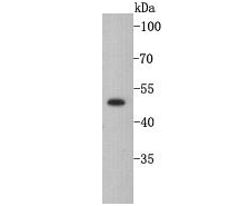
| Molecular Weight(MW) | 51 kDa |
| Cellular Localization | Cytoplasm |
| SwissProt ID | P08729 |
|---|
Application
Fig1: Western blot analysis of Cytokeratin 7 on A549 cell lysate. Proteins were transferred to a PVDF membrane and blocked with 5% BSA in PBS for 1 hour at room temperature. The primary antibody was used at a 1:500 dilution in 5% BSA at room temperature for 2 hours. Goat Anti-Rabbit IgG - HRP Secondary Antibody (HA1001) at 1:5,000 dilution was used for 1 hour at room temperature.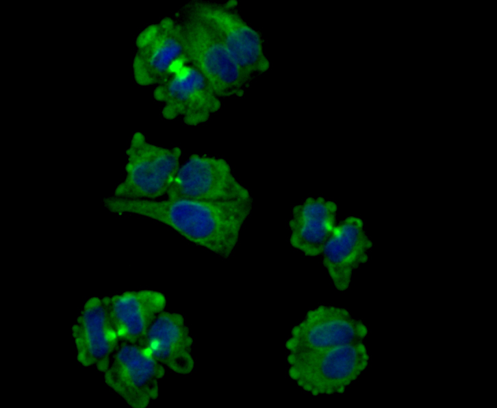
Application
Fig2: ICC staining Cytokeratin 7 in Hela cells (green). Formalin fixed cells were permeabilized with 0.1% Triton X-100 in TBS for 10 minutes at room temperature and blocked with 1% Blocker BSA for 15 minutes at room temperature. Cells were probed with Cytokeratin 7 polyclonal antibody at a dilution of 1:100 for 1 hour at room temperature, washed with PBS. AlexaFluor®488 Goat anti-Rabbit IgG was used as the secondary antibody at 1/100 dilution. The nuclear counter stain is DAPI (blue).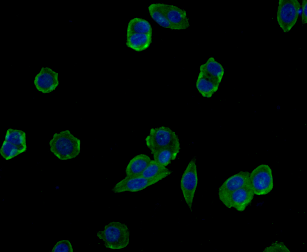
Application
Fig3: ICC staining Cytokeratin 7 in MCF-7 cells (green). Formalin fixed cells were permeabilized with 0.1% Triton X-100 in TBS for 10 minutes at room temperature and blocked with 1% Blocker BSA for 15 minutes at room temperature. Cells were probed with Cytokeratin 7 polyclonal antibody at a dilution of 1:100 for 1 hour at room temperature, washed with PBS. AlexaFluor®488 Goat anti-Rabbit IgG was used as the secondary antibody at 1/100 dilution. The nuclear counter stain is DAPI (blue).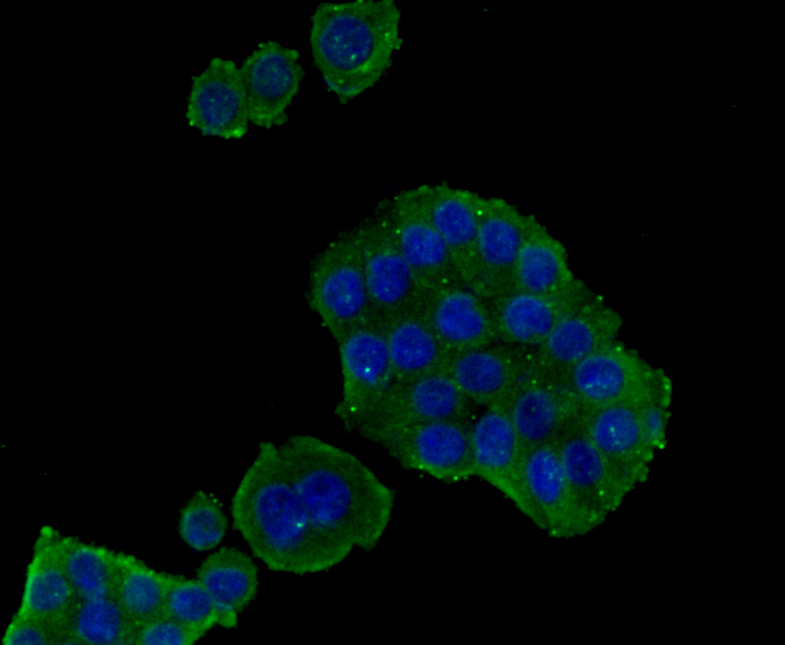
Application
Fig4: ICC staining Cytokeratin 7 in SW480 cells (green). Formalin fixed cells were permeabilized with 0.1% Triton X-100 in TBS for 10 minutes at room temperature and blocked with 1% Blocker BSA for 15 minutes at room temperature. Cells were probed with Cytokeratin 7 polyclonal antibody at a dilution of 1:100 for 1 hour at room temperature, washed with PBS. AlexaFluor®488 Goat anti-Rabbit IgG was used as the secondary antibody at 1/100 dilution. The nuclear counter stain is DAPI (blue).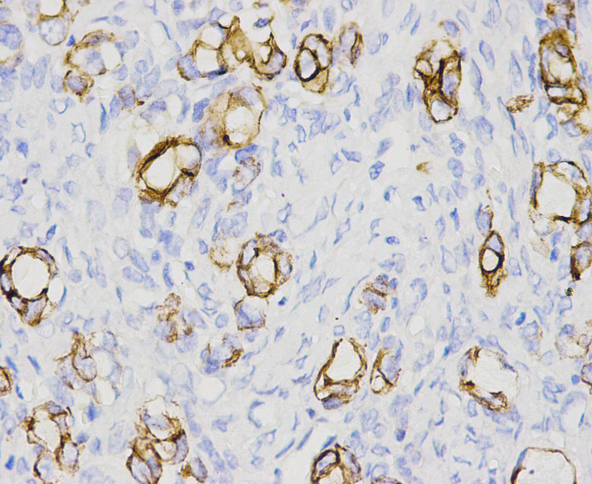
Application
Fig5: Immunohistochemical analysis of paraffin-embedded human ovary carcinoma tissue using anti-Cytokeratin 7 antibody. The section was pre-treated using heat mediated antigen retrieval with Tris-EDTA buffer (pH 8.0-8.4) for 20 minutes.The tissues were blocked in 5% BSA for 30 minutes at room temperature, washed with ddH2O and PBS, and then probed with R1309-4 at 1/100 dilution, for 30 minutes at room temperature and detected using an HRP conjugated compact polymer system. DAB was used as the chrogen. Counter stained with hematoxylin and mounted with DPX.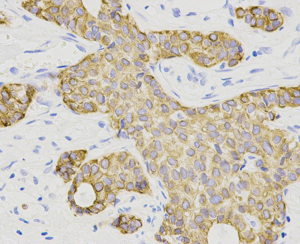
Application
Fig6: Immunohistochemical analysis of paraffin-embedded human breast carcinoma tissue using anti-Cytokeratin 7 antibody. The section was pre-treated using heat mediated antigen retrieval with Tris-EDTA buffer (pH 8.0-8.4) for 20 minutes.The tissues were blocked in 5% BSA for 30 minutes at room temperature, washed with ddH2O and PBS, and then probed with R1309-4 at 1/100 dilution, for 30 minutes at room temperature and detected using an HRP conjugated compact polymer system. DAB was used as the chrogen. Counter stained with hematoxylin and mounted with DPX.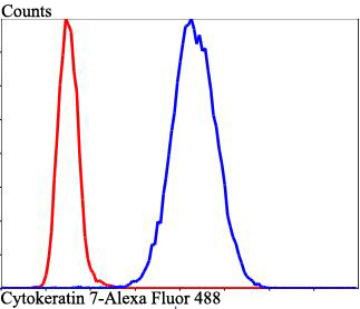
Application
Fig7: Flow cytometric analysis of Cytokeratin 7 was done on Hela cells. The cells were fixed, permeabilized and stained with Cytokeratin 7 antibody at 1/100 dilution (red) compared with an unlabelled control (cells without incubation with primary antibody; blue). After incubation of the primary antibody on room temperature for an hour, the cells was stained with a Alexa Fluor 488-conjugated goat anti-rabbit IgG Secondary antibody at 1/500 dilution for at least 30 minutes.| Positive Control | A549, Hela, MCF-7, SW480, human breast carcinoma tissue, human ovary carcinoma tissue. |
|---|---|
| Application Notes | WB 1:500: ICC 1:200: IHC 1:200: FC 1:50-1:100: |
| Form | Liquid |
|---|---|
| Storage Instructions | Store at +4℃ after thawing. Aliquot store at -20℃. Avoid repeated freeze / thaw cycles. |
| Storage Buffer | 1*TBS (pH7.4), 1%BSA, 40%Glycerol. Preservative: 0.05% Sodium Azide. |
抱歉,暂无相关文献







