| 存储条件 |
|---|
Application
Fig1: Western blot analysis on Hela cell lysates using anti-CHFR rabbit polyclonal antibodies.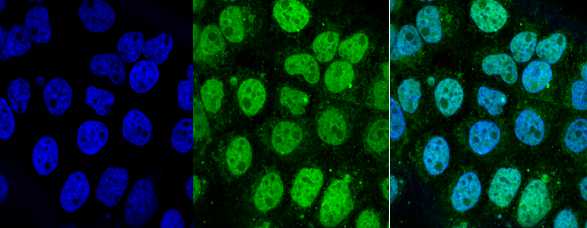
Application
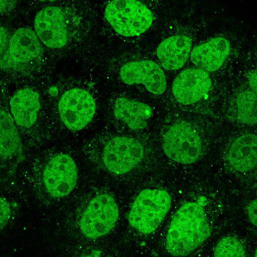
Fig2: ICC staining CHFR in A431 cells (green). Cells were fixed in paraformaldehyde, permeabilised with 0.25% Triton X100/PBS and counterstained with DAPI in order to highlight the nucleus (blue).抱歉,暂无相关文献







