Anti-beta Actin抗体
参阅全部 beta Actin 一抗
兔多克隆抗体to beta Actin
Rabbit
From Jan 2024, QC testing of replenishment batches of this polyclonal changed. All tested and expected application and reactive species combinations are still covered by our Abcam product promise. However, we no longer test all applications. For more information on a specific batch, please contact our Scientific Support who will be happy to help. The immunogen used for this product shares 77% homology with Gamma actin/actin cytoplasmic 2. Cross-reactivity with this protein has not been confirmed experimentally.
适用于: WB, IHC-P, ICC/IFmore details
与反应: Mouse, Rat, Rabbit, Chicken, Cow, Dog, Human, Xenopus laevis, Fish, Chinese hamster
预测可用于: Sheep, Guinea pig, Pig, Drosophila melanogaster, Monkey, Zebrafish, Rhesus monkey![]()
Synthetic peptide. This information is proprietary to Abcam and/or its suppliers.
WB: A431, HeLa, Jurkat, HEK-293, NIH/3T3, MDCK, EBTr, SL-29, CHO and PC-12 whole cell lysate. Rat liver tissue lysate. HeLa nuclear lysate. Fish and rabbit liver. Xenopus laevis embryo. ICC: SV40LT-SMC and NIH/3T3 cells. IHC-P: Rat small intestine tissue. Human colon tissue.
For western blot, milk blocking is suitable for use with fluorescent detection systems. For western blot using chemiluminescent (ECL) systems we recommend BSA blocking.
Abcam recommended secondaries - Goat Anti-Rabbit HRP (ab205718) and Goat Anti-Rabbit Alexa Fluor® 488 (ab150077). Or search our wide range of secondary antibodies for use with your experiment.
The Life Science industry has been in the grips of a reproducibility crisis for a number of years. Abcam is leading the way in addressing this with our range of recombinant monoclonal antibodies and knockout edited cell lines for gold-standard validation. Please check that this product meets your needs before purchasing.
If you have any questions, special requirements or concerns, please send us an inquiry and/or contact our Support team ahead of purchase. Recommended alternatives for this product can be found below, along with publications, customer reviews and Q&As
Liquid
Shipped at 4°C. Store at +4°C short term (1-2 weeks). Upon delivery aliquot. Store at -20°C or -80°C. Avoid freeze / thaw cycle.
pH: 7.40
Preservative: 0.02% Sodium azide
Constituents: 1% BSA, 98.98% PBS
Batches of this product that have a concentration < 1mg/ml may have BSA added as a stabilising agent. If you would like information about the formulation of a specific lot, please contact our scientific support team who will be happy to help.
浓度
批次浓度范围 50 µg 浓度为 0.3 - 1 mg/ml
Immunogen affinity purified
多克隆
IgG
Abpromise™承诺保证使用ab8227于以下的经测试应用
“应用说明”部分 下显示的仅为推荐的起始稀释度;实际最佳的稀释度/浓度应由使用者检定。
| 应用 | Ab评论 | 说明 |
|---|---|---|
| WB | (83) | 1/1000 - 1/5000. We recommend Goat Anti-Rabbit IgG H&L (HRP) (ab6721) secondary antibody. A stronger signal is observed in chemiluminescent western blot when using 2% BSA as the blocking agent. Milk blocking is suitable for use with fluorescent detection systems. Read More |
| IHC-P | (2) | Use a concentration of 0.2 - 1 µg/ml. Perform heat mediated antigen retrieval before commencing with IHC staining protocol. We recommend Goat Anti-Rabbit IgG H&L (Biotin) (ab6720) secondary antibody or Goat Anti-Rabbit IgG Fc (HRP) (ab97200) secondary antibody. Read More |
| ICC/IF | (5) | Use a concentration of 1 µg/ml. |
Entrez Gene: 396526 Chicken
Entrez Gene: 60 Human
Entrez Gene: 11461 Mouse
Entrez Gene: 100009272 Rabbit
Entrez Gene: 574285 Rhesus monkey
Entrez Gene: 443052 Sheep
Entrez Gene: 398459 Xenopus laevis
Entrez Gene: 57934 Zebrafish
Omim: 102630 Human
SwissProt: P60706 Chicken
SwissProt: P48975 Chinese hamster
SwissProt: P60709 Human
SwissProt: P60710 Mouse
SwissProt: P29751 Rabbit
SwissProt: O93400 Xenopus laevis
SwissProt: Q7ZVI7 Zebrafish
Unigene: 520640 Human
Unigene: 708120 Human
Unigene: 727576 Human
Unigene: 328431 Mouse
Unigene: 391967 Mouse
Unigene: 94978 Rat
Unigene: 4138 Xenopus laevis
Unigene: 35143 Zebrafish
A26C1A antibody
A26C1B antibody
ACTB antibody
ACTB_HUMAN antibody
Actin beta antibody
Actin cytoplasmic 1 antibody
Actin, cytoplasmic 1, N-terminally processed antibody
Actx antibody
b actin antibody
b-actin antibody
Beta cytoskeletal actin antibody
Beta-actin antibody
BRWS1 antibody
E430023M04Rik antibody
Melanoma X actin antibody
MGC128179 antibody
PS1TP5 binding protein 1 antibody
PS1TP5BP1 antibody

Immunohistochemistry (Formalin/PFA-fixed paraffin-embedded sections) - Anti-beta Actin antibody (ab8227)
Immunohistochemical analysis of formalin fixed paraffin embedded human colon labelling beta actin with ab8227 at a concentration of 2µg/ml. The immunostaining was performed on a Leica Biosystems BOND® RX instrument with a Bond™ Polymer Refine Detection kit. Heat mediated antigen retrieval was performed with Tris-EDTA buffer (pH 9.0, Epitope Retrieval Solution 2) for 20 mins. ab8227 anti-beta actin antibody was incubated for 30 mins at room temperature. Sections were counterstained with Hematoxylin. Image inset shows absence of staining in secondary antibody only control.

Immunohistochemistry (Formalin/PFA-fixed paraffin-embedded sections) - Anti-beta Actin antibody (ab8227)
Immunohistochemical analysis of formalin fixed paraffin embedded human colon labelling beta actin with ab8227 at a concentration of 1µg/ml. The immunostaining was performed on a Ventana DISCOVERY ULTRA (Roche Tissue Diagnostics) instrument with an OptiView DAB IHC Detection Kit. Heat mediated antigen retrieval was conducted for 32 mins at 100°C with ULTRA cell conditioning solution (CC1, pH 8.5). ab8227 anti-beta actin antibody was incubated at 37°C for 16 mins. Sections were counterstained with Hematoxylin II. Image inset shows absence of staining in secondary antibody only control.

Western blot - Anti-beta Actin antibody (ab8227)
All lanes : Anti-beta Actin antibody (ab8227) at 1/1000 dilution
Lane 1 : A431 (human epidermoid carcinoma cell line) Whole Cell Lysate
Lane 2 : HEK-293 (human epithelial cell line from embryonic kidney) Whole Cell Lysate
Lane 3 : NIH/3T3 (mouse embryo fibroblast cell line) Whole Cell Lysate
Lane 4 : PC-12 (rat adrenal gland pheochromocytoma cell line) Whole Cell Lysate
Lysates/proteins at 20 µg per lane.
Secondary
All lanes : Goat Anti-Rabbit IgG H&L (Alexa Fluor® 790) (ab175781) at 1/10000 dilution
Observed band size: 42 kDawhy is the actual band size different from the predicted?
This blot was produced using a 4-12% Bis-tris gel under the MOPS buffer system. The gel was run at 200V for 50 minutes before being transferred onto a nitrocellulose membrane at 30V for 70 minutes. The membrane was then blocked for an hour using 5% Milk before being incubated with ab8227 overnight at 4°C. Antibody binding was detected using Goat Anti-Rabbit IgG H&L (Alexa Fluor® 790) (ab175781) secondary antibody at a 1:10,000 dilution for 1hr at room temperature and then imaged using the Licor Odyssey CLx.

Immunocytochemistry/ Immunofluorescence - Anti-beta Actin antibody (ab8227)
ab8227 staining beta Actin in SV40LT-SMC (rat aortic smooth muscle cells transfected with SV40). The cells were fixed with 4% formaldehyde (10min), permeabilized with 0.1% Triton X-100 for 5 minutes and then blocked with 1% BSA/10% normal goat serum/0.3M glycine in 0.1%PBS-Tween for 1h. The cells were then incubated overnight at +4°C with ab8227 at 1μg/ml (detected with ab150081, Alexa Fluor® 488 Goat anti-Rabbit, 1μg/ml, shown in green); and ab195889, Mouse monoclonal [DM1A] to alpha Tubulin - Microtubule Marker (Alexa Fluor® 594), at 1/250 dilution (shown in red). Nuclear DNA was labelled with DAPI (shown in blue).
Image was taken with a confocal microscope (Leica-Microsystems, TCS SP8).

Immunohistochemistry (Formalin/PFA-fixed paraffin-embedded sections) - Anti-beta Actin antibody (ab8227)
IHC image of ab8227 staining beta Actin in rat small intestine formalin fixed paraffin embedded tissue sections, performed on a Leica Bond. The section was pre-treated using heat mediated antigen retrieval with EDTA (epitope retrieval solution 2) for 20 mins. The section was then incubated with ab8227, 0.2μg/ml, for 15 mins at room temperature and detected using an HRP conjugated compact polymer system. DAB was used as the chromogen. The section was then counterstained with haematoxylin and mounted with DPX. No primary antibody was used in the secondary only control (shown on the inset).
For other IHC staining systems (automated and non-automated) customers should optimize variable parameters such as antigen retrieval conditions, primary antibody concentration and antibody incubation times.

Immunocytochemistry/ Immunofluorescence - Anti-beta Actin antibody (ab8227)
ab8227 staining beta Actin in NIH/3T3 (mouse embryo fibroblast cell line) cells. The cells were fixed with 100% methanol (5min), permeabilized with 0.1% Triton X-100 for 5 minutes and then blocked with 1% BSA/10% normal goat serum/0.3M glycine in 0.1%PBS-Tween for 1h. The cells were then incubated overnight at +4°C with ab8227 at 1μg/ml (shown in green) and ab195889, Mouse monoclonal [DM1A] to alpha Tubulin - Microtubule Marker (Alexa Fluor® 594), at 1/250 dilution (shown in red). Nuclear DNA was labelled with DAPI (shown in blue).
Image was taken with a confocal microscope (Leica-Microsystems, TCS SP8).

Immunohistochemistry (Formalin/PFA-fixed paraffin-embedded sections) - Anti-beta Actin antibody (ab8227)
IHC image of beta actin staining in a section of formalin-fixed paraffin-embedded normal human colon*. The section was pre-treated using pressure cooker heat mediated antigen retrieval with sodium citrate buffer (pH6) for 30mins. The section was then incubated with ab8227, 1/1000 dilution, for 15 mins at room temperature. A goat anti-rabbit biotinylated secondary antibody (ab6720, 1/1000 dilution) was used to detect the primary, and visualized using an HRP conjugated ABC system. Streptavidin HRP was used, ab7403 at a 1/10000 dilution. DAB was used as the chromogen (ab103723), diluted 1/100 and incubated for 10min at room temperature. The section was then counterstained with haematoxylin and mounted with DPX.
The inset negative control image is taken from an identical assay without primary antibody.
For other IHC staining systems (automated and non-automated) customers should optimize variable parameters such as antigen retrieval conditions, primary antibody concentration and antibody incubation times.
*Tissue obtained from the Human Research Tissue Bank, supported by the NIHR Cambridge Biomedical Research Centre

Western blot - Anti-beta Actin antibody (ab8227)
All lanes : Anti-beta Actin antibody (ab8227) at 1 µg/ml
Lane 1 : HeLa (human epithelial cell line from cervix adenocarcinoma) whole cell lysate (blocked with 2% BSA)
Lane 2 : NIH/3T3 (mouse embryo fibroblast cell line) whole cell lysate (blocked with 2% BSA)
Lane 3 : Rat Liver tissue lysate (blocked with 2% BSA)
Lane 4 : HeLa whole cell lysate (blocked with 3% Milk)
Lane 5 : NIH/3T3 whole cell lysate (blocked with 3% Milk)
Lane 6 : Rat Liver tissue lysate (blocked with 3% Milk)
Lysates/proteins at 10 µg per lane.
Secondary
All lanes : Goat Anti-Rabbit IgG H&L (HRP) at 1/50000 dilution
Developed using the ECL technique.
Performed under reducing conditions.
Observed band size: 45 kDawhy is the actual band size different from the predicted?
Exposure time: 10 seconds
Lanes 1-3: Blocked with 2% BSA
Lanes 4-6: Blocked with 3% Milk
This blot was produced using a 4-12% Bis-tris gel under the MOPS buffer system. The gel was run at 200V for 50 minutes before being transferred onto a Nitrocellulose membrane at 30V for 70 minutes. The membrane was then blocked for an hour using 2% Bovine Serum Albumin or 3% Milk before being incubated with ab8227 overnight at 4°C. Antibody binding was detected using an anti rabbit HRP secondary antibody, and visualised using ECL development solution ab133406

Western blot - Anti-beta Actin antibody (ab8227)
All lanes : Anti-beta Actin antibody (ab8227) at 0.1 µg/ml
Lane 1 : HeLa (human epithelial cell line from cervix adenocarcinoma) Whole Cell Lysate
Lane 2 : NIH/3T3 (mouse embryonic fibroblast cell line) Whole Cell Lysate
Lane 3 : Liver (Rat) Tissue Lysate
Lysates/proteins at 10 µg per lane.
Secondary
All lanes : Goat Anti-Rabbit IgG H&L (HRP) (ab97051) at 1/50000 dilution
Performed under reducing conditions.
Observed band size: 42 kDawhy is the actual band size different from the predicted?
Exposure time: 1 minute
This blot was produced using a 4-12% Bis-tris gel under the MOPS buffer system. The gel was run at 200V for 50 minutes before being transferred onto a Nitrocellulose membrane at 30V for 70 minutes. The membrane was then blocked for an hour using 2% Bovine Serum Albumin before being incubated with ab8227 overnight at 4°C. Antibody binding was detected using an anti-rabbit HRP secondary antibody, and visualised using ECL development solution ab133406
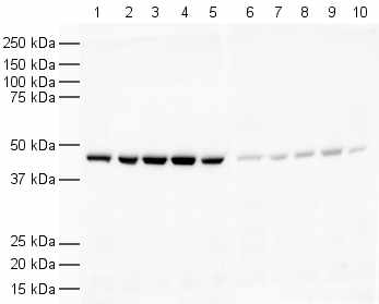
Western blot - Anti-beta Actin antibody (ab8227)
All lanes : Anti-beta Actin antibody (ab8227) at 1/1000 dilution
Lane 1 : HeLa (human epithelial cell line from cervix adenocarcinoma) nuclear lysate
Lane 2 : HeLa whole cell lysate
Lane 3 : A431 (human epidermoid carcinoma cell line) cell lysate
Lane 4 : Jurkat (human T cell leukemia cell line from peripheral blood) cell lysate
Lane 5 : HEK-293 (human epithelial cell line from embryonic kidney) cell lysate
Lane 6 : HeLa nuclear lysate with Human beta Actin peptide (ab13772) at 1 µg/ml
Lane 7 : HeLa whole cell lysate with Human beta Actin peptide (ab13772) at 1 µg/ml
Lane 8 : A431 cell lysate with Human beta Actin peptide (ab13772) at 1 µg/ml
Lane 9 : Jurkat cell lysate with Human beta Actin peptide (ab13772) at 1 µg/ml
Lane 10 : HEK-293 cell lysate with Human beta Actin peptide (ab13772) at 1 µg/ml
Lysates/proteins at 20 µg per lane.
Secondary
All lanes : Goat Anti-Rabbit IgG H&L (HRP) (ab6721) at 1/5000 dilution
Observed band size: 41.7 kDawhy is the actual band size different from the predicted?
Exposure time: 5 seconds

Western blot - Anti-beta Actin antibody (ab8227)
All lanes : Anti-beta Actin antibody (ab8227) at 1/1000 dilution
Lane 1 : HeLa (Human epithelial cell line from cervix adenocarcinoma) cells
Lane 2 : NIH/3T3 (Mouse embryonic fibroblast cell line) cells
Lane 3 : Fish Liver
Lane 4 : Rabbit Liver
Lane 5 : MDCK (Canine kidney cell line) cells
Lane 6 : EBTr (cow trachea) cells
Lane 7 : SL-29 (chicken day 11 embryo) cells
Lane 8 : CHO (Chinese hamster ovary cell line) cells
Lane 9 : Xenopus laevis embryo
Lysates/proteins at 20 µg per lane.
Secondary
All lanes : Goat Anti-Rabbit IgG H&L (HRP) (ab6721) at 1/5000 dilution
Observed band size: 41.7 kDawhy is the actual band size different from the predicted?
Exposure time: 30 seconds

Immunohistochemistry (Formalin/PFA-fixed paraffin-embedded sections) - Anti-beta Actin antibody (ab8227)
IHC image of beta Actin staining in normal human colon, formalin-fixed and paraffin-embedded tissue*. The section was pre-treated using pressure cooker heat mediated antigen retrieval with sodium citrate buffer (pH6) for 30mins. The section was incubated with ab8227, 3µg/ml overnight at +4°C. A anti-rabbit HRP secondary antibody (Ab97200, 1/200 dilution) was used for 1hr at room temperature. The section was counterstained with haematoxylin and mounted with DPX.
The inset negative control image is taken from an identical assay without primary antibody.
*Tissue obtained from the Human Research Tissue Bank, supported by the NIHR Cambridge Biomedical Research Centre


