Anti-Iba1抗体
参阅全部 Iba1 一抗
山羊多克隆抗体to Iba1
Goat
ab5076 is expected to recognize the isoforms represented by NP_001614 and NP_116573 but not NP_004838.
Although we received several excellent papers on ab5076 working with rat IHC-P, rat is not a batch tested species for IHC-P and we cannot guarantee.
适用于: IHC-P, WBmore details
与反应: Rat, Human
预测可用于: Pig, Macaque monkey![]()
Synthetic peptide corresponding to Human Iba1 aa 135-147 (C terminal). Accession Number(s): NP_116573.1; NP_001614.3
Sequence:
C-TGPPAKKAISELP
WB: Human lymph node and rat brain tissue lysate. IHC-P: Human spleen tissue.
The Iba1 antibody (ab5076) is commonly used as a marker of microglia activation in staining and immunohistochemistry, given that ionized calcium binding adaptor molecule 1 (Iba1) is a microglia/macrophage-specific calcium-binding protein with actin-bundling activity that participates in membrane ruffling and phagocytosis in activated microglia.
The Life Science industry has been in the grips of a reproducibility crisis for a number of years. Abcam is leading the way in addressing this with our range of recombinant monoclonal antibodies and knockout edited cell lines for gold-standard validation. Please check that this product meets your needs before purchasing.
If you have any questions, special requirements or concerns, please send us an inquiry and/or contact our Support team ahead of purchase. Recommended alternatives for this product can be found below, along with publications, customer reviews and Q&As
Liquid
Shipped at 4°C. Upon delivery aliquot and store at -20°C. Avoid freeze / thaw cycles.
pH: 7.30
Preservative: 0.02% Sodium azide
Constituents: Tris buffered saline, 0.5% BSA
Immunogen affinity purified
Purified from goat serum by ammonium sulphate precipitation followed by antigen affinity chromatography using the immunizing peptide.
多克隆
IgG
Abpromise™承诺保证使用ab5076于以下的经测试应用
“应用说明”部分 下显示的仅为推荐的起始稀释度;实际最佳的稀释度/浓度应由使用者检定。
| 应用 | Ab评论 | 说明 |
|---|---|---|
| IHC-P | (28) | Use a concentration of 5 µg/ml. Perform heat mediated antigen retrieval with citrate buffer pH 6 before commencing with IHC staining protocol. Although we received several excellent papers on ab5076 working with rat IHC-P, rat is not a batch tested species for IHC-P and we cannot guarantee. Read More |
| WB | (7) | Use a concentration of 1 - 3 µg/ml. Detects a band of approximately 16-17 kDa (predicted molecular weight: 17 kDa). A 1 hour primary incubation is recommended for this product. We received several excellent Abreviews on this antibody working with mouse, however, mouse is not a batch tested species and we cannot guarantee that this antibody will work on mouse. Some of our customers have observed high background in mouse samples. We recommend blocking in milk (3% milk in TBST) for 1 hour at room temperature. Blocking with BSA gives high background. Read More |
Entrez Gene: 199 Human
Omim: 601833 Human
SwissProt: P55008 Human
Unigene: 76364 Human
Unigene: 32080 Rat
AIF 1 antibody
AIF-1 antibody
Aif1 antibody
AIF1 protein antibody
AIF1_HUMAN antibody
Allograft inflammatory factor 1 antibody
Allograft inflammatory factor 1 splice variant G antibody
allograft inflammatory factor-1 splice variant Hara-1 antibody
balloon angioplasty responsive transcription antibody
BART 1 antibody
G1 antibody
G1 putative splice variant of allograft inflamatory factor 1 antibody
IBA 1 antibody
IBA1 antibody
interferon gamma responsive transcript antibody
Interferon responsive transcript 1 antibody
interferon responsive transcript factor 1 antibody
Ionized calcium binding adapter molecule 1 antibody
Ionized calcium-binding adapter molecule 1 antibody
ionized calcium-binding adapter molecule antibody
IRT 1 antibody
IRT1 antibody
Microglia response factor antibody
MRF1 antibody
Protein g1 antibody

Immunohistochemistry (Formalin/PFA-fixed paraffin-embedded sections) - Anti-Iba1 antibody (ab5076)
Immunohistochemistry (Formalin/PFA-fixed paraffin-embedded sections) analysis of human spleen tissue labelling Iba1 with ab5076 at 8µg/ml.
Heat induced antigen retrieval with citrate buffer Ph 6, HRP-staining.

Immunohistochemistry (Formalin/PFA-fixed paraffin-embedded sections) - Anti-Iba1 antibody (ab5076)
Negative control: Immunohistochemistry (Formalin/PFA-fixed paraffin-embedded sections) analysis of human spleen tissue labelling Iba1 with no primary antibody.
Heat induced antigen retrieval with citrate buffer Ph 6, HRP-staining.
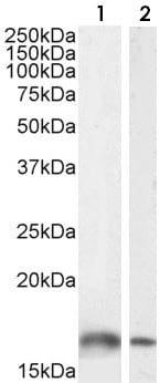
Western blot - Anti-Iba1 antibody (ab5076)
All lanes : Anti-Iba1 antibody (ab5076) at 1 µg/ml
Lane 1 : Human lymph node tissue lysate in RIPA buffer
Lane 2 : Rat brain tissue lysate in RIPA buffer
Lysates/proteins at 35 µg per lane.
Developed using the ECL technique.
Predicted band size: 17 kDa
Detected by chemiluminescence.

Immunohistochemistry (Formalin/PFA-fixed paraffin-embedded sections) - Anti-Iba1 antibody (ab5076)Image from Iris Margalit Trutzer et al, ACTA neuropathologica communications, 7, 40, Fig. 7; doi: 10.1186/s40478-019-0684-8
Immunohistochemical analysis of formalin-fixed, paraffin-embedded human lateral prefrontal cortex tissue labeling iba1 labeled microglia (labeled with black arrows) within the cortex using ab5076 at 1/1000 dilution. (Cropped image).
Sections were rinsed in 0.01 m PBS, pH 7.4, followed by 10% serum matching the species of the secondary antibody, 5% bovine serum albumin, and 0.1% Triton X-100 in 0.01 m PBS blocking solution for 1 h and incubated for 2 days at 4 °C in primary antibody. The sections were rinsed in PBS and incubated overnight at 4 °C with a horse anti-Goat IgG (Biotinylated) secondary antibody.


