| 存储条件 |
|---|
| Product Name | PKC alpha Recombinant Rabbit Monoclonal Antibody |
|---|---|
| Antibody Type | Primary Antibodies |
| Product description | Members of the protein kinase C (PKC) family play a key regulatory role in a variety of cellular functions including cell growth and differentiation, gene expression, hormone secretion and membrane function. PKCs were originally identified as serine/threonine protein kinases whose activity was dependent on calcium and phospholipids. Diacylglycerols (DAG) and tumor-promoting phorbol esters bind to and activate PKC. PKCs can be subdivided into many different isoforms. Patterns of expression for each PKC isoform differ among tissues and PKC family members exhibit clear differences in their cofactor dependencies. For instance, the kinase activities of PKC and |
| Immunogen | Recombinant protein within Human PKC alpha aa 560-672 / 672. |
| Clonality | monoclonal |
|---|---|
| Isotype | IgG |
| Host Species | Recombinant rabbit |
| Tested Applications | FCICC/IFIPWB |
| WB:1:1000-1:2000 ICC/IF:1:50-1:200 IP:1:10-1:50 FC:1:50-1:100 | |
| Species Reactivity | HumanMouseRat |
| Concentration | 1mg/ml |
| Purification | Protein A |
| Alternative Names | AAG6 antibody Aging associated gene 6 antibody aPKC antibody KPCA_HUMAN antibody PKC alpha antibody PKC-A antibody PKC-alpha antibody PKCA antibody PRKACA antibody PRKCA antibody AAG6 antibody Aging associated gene 6 antibody aPKC antibody KPCA_HUMAN antibody PKC alpha antibody PKC-A antibody PKC-alpha antibody PKCA antibody PRKACA antibody PRKCA antibody Protein Kinase C alpha antibody Protein kinase C alpha type antibody |
|---|---|
| Molecular Weight(MW) | 77kDa |
| Cellular Localization | Cytoplasm, Nucleus, Cell membrane, Mitochondrion membrane. |
WB
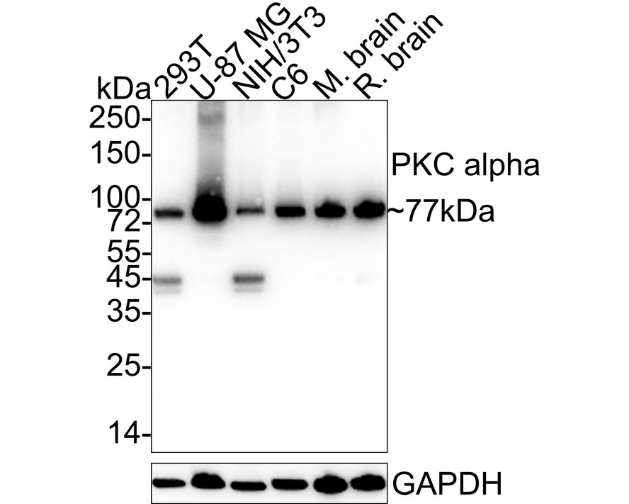
Western blot analysis of PKC alpha on different lysates with Rabbit anti-PKC alpha antibody at 1/1,000 dilution. Lane 1: 293T cell lysate (20 µg/Lane), Lane 2: U-87 MG cell lysate (20 µg/Lane), Lane 3: NIH/3T3 cell lysate (20 µg/Lane), Lane 4: C6 cell lysate (20 µg/Lane), Lane 5: Mouse brain tissue lysate (40 µg/Lane), Lane 6: Rat brain tissue lysate (40 µg/Lane), Exposure time: 9 seconds; 4-20% SDS-PAGE gel. Proteins were transferred to a PVDF membrane and blocked with 5% NFDM/TBST for 1 hour at room temperature. The primary antibody at 1/1,000 dilution was used in 5% NFDM/TBST at 4℃ overnight. Goat Anti-Rabbit IgG - HRP Secondary Antibody at 1/50,000 dilution was used for 1 hour at room temperature.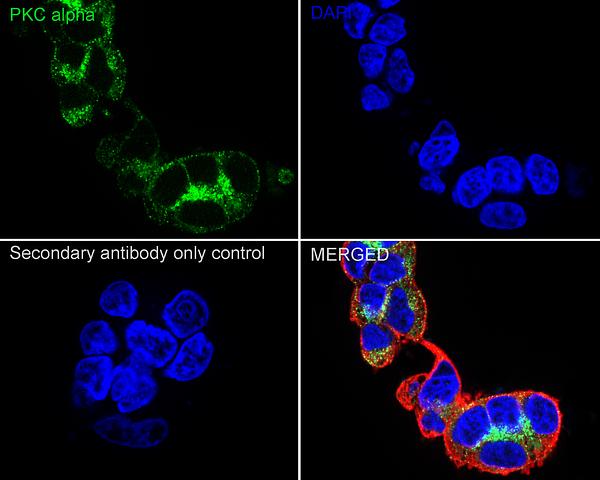
ICC/IF
Immunocytochemistry analysis of 293T cells labeling PKC alpha with Rabbit anti-PKC alpha antibody at 1/100 dilution. Cells were fixed in 4% paraformaldehyde for 15 minutes at room temperature, permeabilized with 0.1% Triton X-100 in PBS for 15 minutes at room temperature, then blocked with 1% BSA in 10% negative goat serum for 1 hour at room temperature. Cells were then incubated with Rabbit anti-PKC alpha antibody at 1/100 dilution in 1% BSA in PBST overnight at 4 ℃. Goat Anti-Rabbit IgG H&L (iFluor™ 488) was used as the secondary antibody at 1/1,000 dilution. PBS instead of the primary antibody was used as the secondary antibody only control. Nuclear DNA was labelled in blue with DAPI. Beta tubulin (red) was stained at 1/100 dilution overnight at +4℃. Goat Anti-Mouse IgG H&L (iFluor™ 594) was used as the secondary antibody at 1/1,000 dilution.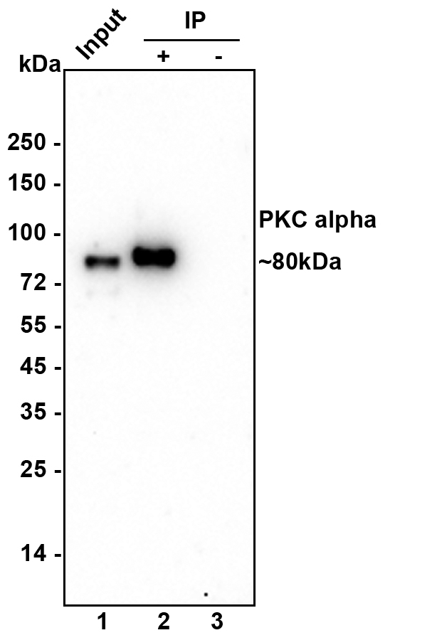
IP
PKC alpha was immunoprecipitated from 0.2 mg 293T cell lysate with Rabbit anti-PKC alpha antibody at 2 µg/10 µl beads. Western blot was performed from the immunoprecipitate using Rabbit anti-PKC alpha antibody at 1/1,000 dilution. Anti-Rabbit IgG for IP Nano-secondary antibody at 1/5,000 dilution was used for 1 hour at room temperature. Lane 1: 293T cell lysate (input) Lane 2: Rabbit anti-PKC alpha antibody IP in 293T cell lysate Lane 3: Rabbit IgG instead of Rabbit anti-PKC alpha antibody in 293T cell lysate Blocking/Dilution buffer: 5% NFDM/TBST Exposure time: 2 min.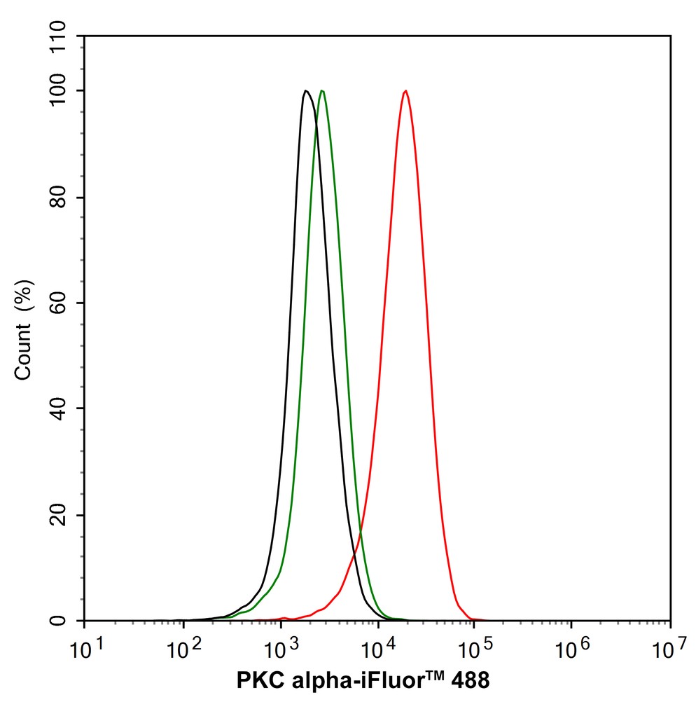
FC
Flow cytometric analysis of 293T cells labeling PKC alpha. Cells were fixed and permeabilized. Then stained with the primary antibody (1/1,000) (red) compared with Rabbit IgG Isotype Control (green). After incubation of the primary antibody at +4℃ for an hour, the cells were stained with a iFluor™ 488 conjugate-Goat anti-Rabbit IgG Secondary antibody at 1/1,000 dilution for 30 minutes at +4℃. Unlabelled sample was used as a control (cells without incubation with primary antibody; black).| Positive Control | K562, MCF-7, Hela, CRC, A549, PC12, mouse brain tissue, mouse small intestine tissue, mouse heart tissue, human lung tissue. |
|---|---|
| Application Notes | WB:1:1000-1:2000 ICC/IF:1:50-1:200 IP:1:10-1:50 FC:1:50-1:100 |
| Form | Liquid |
|---|---|
| Storage Instructions | Store at +4℃ after thawing. Aliquot store at -20℃ or -80℃. Avoid repeated freeze / thaw cycles. |
| Storage Buffer | 1*TBS (pH7.4), 1%BSA, 40%Glycerol. Preservative: 0.05% Sodium Azide. |
抱歉,暂无相关文献







