Anti-CREB + ICER抗体
兔多克隆抗体to CREB + ICER
Rabbit
ab5803 detects both the phosphorylated and non-phosphorylated forms of cyclic-AMP response element binding protein (CREB) from rat cells.
适用于: WB, ICC/IF, IHC-Pmore details
与反应: Mouse, Rat, Human
预测可用于: Cow, Dog, Zebrafish![]()
Synthetic peptide corresponding to Human CREB aa 123-136.
Sequence:
KRREILSRRPSYRK
(Peptide available as ab5860)
WB: GH4 cell extract. ICC/IF: SK-N-MC cells, Neuro-2a cells. IHC-P: Mouse brain tissue, Human glioma, Human lung adenocarcinoma.
The Life Science industry has been in the grips of a reproducibility crisis for a number of years. Abcam is leading the way in addressing this with our range of recombinant monoclonal antibodies and knockout edited cell lines for gold-standard validation. Please check that this product meets your needs before purchasing.
If you have any questions, special requirements or concerns, please send us an inquiry and/or contact our Support team ahead of purchase. Recommended alternatives for this product can be found below, along with publications, customer reviews and Q&As
Liquid
Shipped at 4°C. Store at +4°C short term (1-2 weeks). Upon delivery aliquot. Store at -20°C or -80°C. Avoid freeze / thaw cycle.
Preservative: 0.05% Sodium azide
Constituents: 0.1% BSA, 99% PBS
浓度
100 µg 浓度为 1 mg/ml
Immunogen affinity purified
多克隆
IgG
Abpromise™承诺保证使用ab5803于以下的经测试应用
“应用说明”部分 下显示的仅为推荐的起始稀释度;实际最佳的稀释度/浓度应由使用者检定。
| 应用 | Ab评论 | 说明 |
|---|---|---|
| WB | (2) | Use a concentration of 2 µg/ml. Detects a band of approximately 43 kDa (predicted molecular weight: 43 kDa).Can be blocked with CREB + ICER peptide (ab5860). |
| ICC/IF | (1) | 1/50 - 1/500. |
| IHC-P | 1/20 - 1/200. |
Entrez Gene: 1385 Human
Entrez Gene: 1390 Human
Entrez Gene: 12912 Mouse
Entrez Gene: 12916 Mouse
Omim: 123810 Human
Omim: 123812 Human
SwissProt: P16220 Human
SwissProt: Q03060 Human
SwissProt: P27699 Mouse
SwissProt: Q01147 Mouse
Unigene: 200250 Human
Unigene: 516646 Human
Unigene: 453295 Mouse
Unigene: 5244 Mouse
Unigene: 10251 Rat
Unigene: 90061 Rat

Western blot - Anti-CREB + ICER antibody (ab5803)
Shows a Western blot of CREB on GH4 cell extract using ab5803.

Immunocytochemistry/ Immunofluorescence - Anti-CREB + ICER antibody (ab5803)
Immunofluorescent analysis of CREB (green) showing staining in the nucleus of SK-N-MC cells (right) compared to a negative control without primary antibody (left). Formalin-fixed cells were permeabilized with 0.1% Triton X-100 in TBS for 5-10 minutes and blocked with 3% BSA-PBS for 30 minutes at room temperature. Cells were probed with a CREB polyclonal antibody (ab5803) in 3% BSA-PBS at a dilution of 1:200 and incubated overnight at 4ºC in a humidified chamber. Cells were washed with PBST and incubated with a DyLight-conjugated secondary antibody in PBS at room temperature in the dark. Actin was stained using Alexa Fluor 554 (red) and nuclei were stained with Hoechst or DAPI (blue). Images were taken at a magnification of 60x.

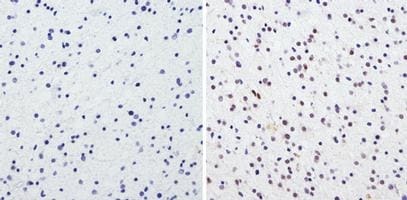
Immunohistochemistry (Formalin/PFA-fixed paraffin-embedded sections) - Anti-CREB + ICER antibody (ab5803)
Immunohistochemistry analysis of CREB showing staining in the nucleus of paraffin-embedded mouse brain tissue (right) compared to a negative control without primary antibody (left). To expose target proteins, antigen retrieval was performed using 10mM sodium citrate (pH 6.0), microwaved for 8-15 min. Following antigen retrieval, tissues were blocked in 3% H2O2-methanol for 15 min at room temperature, washed with ddH2O and PBS, and then probed with a CREB polyclonal antibody (ab5803) diluted in 3% BSA-PBS at a dilution of 1:50 overnight at 4°C in a humidified chamber. Tissues were washed extensively in PBST and detection was performed using an HRP-conjugated secondary antibody followed by colorimetric detection using a DAB kit. Tissues were counterstained with hematoxylin and dehydrated with ethanol and xylene to prep for mounting.
Immunohistochemistry (Formalin/PFA-fixed paraffin-embedded sections) - Anti-CREB + ICER antibody (ab5803)
Immunohistochemistry analysis of CREB showing staining in the nucleus of paraffin-embedded Human glioma (right) compared to a negative control without primary antibody (left). To expose target proteins, antigen retrieval was performed using 10mM sodium citrate (pH 6.0), microwaved for 8-15 min. Following antigen retrieval, tissues were blocked in 3% H2O2-methanol for 15 min at room temperature, washed with ddH2O and PBS, and then probed with a CREB polyclonal antibody (ab5803) diluted in 3% BSA-PBS at a dilution of 1:100 overnight at 4°C in a humidified chamber. Tissues were washed extensively in PBST and detection was performed using an HRP-conjugated secondary antibody followed by colorimetric detection using a DAB kit. Tissues were counterstained with hematoxylin and dehydrated with ethanol and xylene to prep for mounting.

Immunohistochemistry (Formalin/PFA-fixed paraffin-embedded sections) - Anti-CREB + ICER antibody (ab5803)
Immunohistochemistry analysis of CREB showing staining in the nucleus of paraffin-embedded Human lung adenocarcinoma (right) compared to a negative control without primary antibody (left). To expose target proteins, antigen retrieval was performed using 10mM sodium citrate (pH 6.0), microwaved for 8-15 min. Following antigen retrieval, tissues were blocked in 3% H2O2-methanol for 15 min at room temperature, washed with ddH2O and PBS, and then probed with a CREB polyclonal antibody (ab5803) diluted in 3% BSA-PBS at a dilution of 1:100 overnight at 4°C in a humidified chamber. Tissues were washed extensively in PBST and detection was performed using an HRP-conjugated secondary antibody followed by colorimetric detection using a DAB kit. Tissues were counterstained with hematoxylin and dehydrated with ethanol and xylene to prep for mounting.

Immunocytochemistry/ Immunofluorescence - Anti-CREB + ICER antibody (ab5803)
Immunofluorescent analysis of CREB (green) showing staining in the nucleus of Neuro-2a cells (right) compared to a negative control without primary antibody (left). Formalin-fixed cells were permeabilized with 0.1% Triton X-100 in TBS for 5-10 minutes and blocked with 3% BSA-PBS for 30 minutes at room temperature. Cells were probed with a CREB polyclonal antibody (ab5803) in 3% BSA-PBS at a dilution of 1:100 and incubated overnight at 4ºC in a humidified chamber. Cells were washed with PBST and incubated with a DyLight-conjugated secondary antibody in PBS at room temperature in the dark. Nuclei were stained with Hoechst or DAPI (blue). Images were taken at a magnification of 60x.

Western blot - Anti-CREB + ICER antibody (ab5803)This image is courtesy of Richard D'Mello, Kings College, London
All lanes : Anti-CREB + ICER antibody (ab5803) at 1/500 dilution
All lanes : hippocampal lysate
Lysates/proteins at 40 µg per lane.
Secondary
All lanes : Donkey Anti-Rabbit IR800-linked conjugated to IRDye 800CW at 1/15000 dilution
Performed under reducing conditions.
Predicted band size: 43 kDa
Observed band size: 43 kDa
Additional bands at: 55 kDa (possible non-specific binding)
Exposure time: 5 minutes


