Anti-Dynamin 3抗体
参阅全部 Dynamin 3 一抗
兔多克隆抗体to Dynamin 3
Rabbit
适用于: WB, IHC-P, ICC/IFmore details
与反应: Rat, Human
Synthetic peptide corresponding to Rat Dynamin 3 aa 623-639.
Sequence:
PDKSFTENDENGQAENF
(Peptide available as ab4986)
The Life Science industry has been in the grips of a reproducibility crisis for a number of years. Abcam is leading the way in addressing this with our range of recombinant monoclonal antibodies and knockout edited cell lines for gold-standard validation. Please check that this product meets your needs before purchasing.
If you have any questions, special requirements or concerns, please send us an inquiry and/or contact our Support team ahead of purchase. Recommended alternatives for this product can be found below, along with publications, customer reviews and Q&As
Liquid
Shipped at 4°C. Store at +4°C short term (1-2 weeks). Upon delivery aliquot. Store at -20°C or -80°C. Avoid freeze / thaw cycle.
Preservative: 0.05% Sodium azide
Constituents: 0.1% BSA, 99% PBS
Immunogen affinity purified
The dynamins are a family of 100 kDa GTPases transcribed from at least three separate genes. At least four mRNA splice variants for each dynamin have been described. Dynamins contain several conserved regions including the conserved, amino-terminal GTPase domain, a centrally located membrane-binding plekstrin homology domain (PHD), and a coiled-coil region located in front of a proline-rich domain (PRD). The PRD is thought to mediate interactions between dynamin and numerous other cellular proteins. Dynamin 1 is expressed exclusively in neurons, Dynamin 2 is ubiquitously expressed, and Dynamin 3 is thought to be restricted to expression in the brain, testis, heart, and lung. The dynamins participate in the cellular process of clathrin-mediated and fluid-phase endocytosis.
多克隆
IgG
Abpromise™承诺保证使用ab3458于以下的经测试应用
“应用说明”部分 下显示的仅为推荐的起始稀释度;实际最佳的稀释度/浓度应由使用者检定。
| 应用 | Ab评论 | 说明 |
|---|---|---|
| WB | (1) | Use at an assay dependent concentration. By Western blot, this antibody detects an ~100 kDa protein representing Dynamin 3 from HeLa cell lysate. |
| IHC-P | Use a concentration of 4 µg/ml. | |
| ICC/IF | (1) | Use a concentration of 1 µg/ml. |
Entrez Gene: 26052 Human
Omim: 611445 Human
SwissProt: Q9UQ16 Human
Unigene: 654775 Human
Unigene: 11191 Rat
DNM3 antibody
DNM3 protein antibody
Dyn3 antibody
DYN3_HUMAN antibody
Dyna III antibody
DynaIII antibody
dynamin 2 antibody
Dynamin antibody
Dynamin family member antibody
Dynamin testicular antibody
Dynamin-3 antibody
Dynamin3 antibody
EC 3.6.5.5 antibody
KIAA0820 antibody
MGC70433 antibody
T dynamin antibody
T-dynamin antibody
testicular dynamin antibody
testicular antibody
DNM 3 antibody
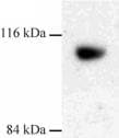
Western blot - Anti-Dynamin 3 antibody (ab3458)
Western blot of Dynamin 3 on rat liver extract using ab3458.

Immunohistochemistry (Formalin/PFA-fixed paraffin-embedded sections) - Anti-Dynamin 3 antibody (ab3458)
ab3458 (4µg/ml) staining Dynamin 3 in human brain cerebellum using an automated system (DAKO Autostainer Plus). Using this protocol there is strong staining of nuclear/cytoplasmic compartments within the white matter region
Sections were rehydrated and antigen retrieved with the Dako 3 in 1 AR buffer citrate pH 6.1 in a DAKO PT link. Slides were peroxidase blocked in 3% H2O2 in methanol for 10 mins. They were then blocked with Dako Protein block for 10 minutes (containing casein 0.25% in PBS) then incubated with primary antibody for 20 min and detected with Dako envision flex amplification kit for 30 minutes. Colorimetric detection was completed with Diaminobenzidine for 5 minutes. Slides were counterstained with Haematoxylin and coverslipped under DePeX. Please note that, for manual staining, optimization of primary antibody concentration and incubation time is recommended. Signal amplification may be required.

Immunocytochemistry/ Immunofluorescence - Anti-Dynamin 3 antibody (ab3458)
ICC/IF image of ab3458 stained HeLa cells. The cells were 100% methanol fixed (5 min) and then incubated in 1%BSA / 10% normal goat serum / 0.3M glycine in 0.1% PBS-Tween for 1h to permeabilise the cells and block non-specific protein-protein interactions. The cells were then incubated with the antibody (ab3458, 1µg/ml) overnight at +4°C. The secondary antibody (green) was Alexa Fluor® 488 goat anti-rabbit IgG (H+L) used at a 1/1000 dilution for 1h. Alexa Fluor® 594 WGA was used to label plasma membranes (red) at a 1/200 dilution for 1h. DAPI was used to stain the cell nuclei (blue) at a concentration of 1.43µM.


