Anti-Proteasome 20S C2/HC2抗体
参阅全部 Proteasome 20S C2/HC2 一抗
兔多克隆抗体to Proteasome 20S C2/HC2
Rabbit
Detects proteasome 20S C2/HC2 subunit.
适用于: WB, IHC-P, ICC/IFmore details
与反应: Mouse, Rat, Hamster, Dog, Human, Chinese hamster
预测可用于: Chicken, Cow, Cynomolgus monkey![]()
Synthetic peptide corresponding to Human Proteasome 20S C2/HC2 aa 249-263 (C terminal).
Sequence:
PADEPAEKADEPMEH
WB: MDA-MB-231, MCF7, PC-3, HepG2 and Jurkat whole cell lysate, CHO whole cell lysate. ICC/IF: MDA-MB-231 cells.
The Life Science industry has been in the grips of a reproducibility crisis for a number of years. Abcam is leading the way in addressing this with our range of recombinant monoclonal antibodies and knockout edited cell lines for gold-standard validation. Please check that this product meets your needs before purchasing.
If you have any questions, special requirements or concerns, please send us an inquiry and/or contact our Support team ahead of purchase. Recommended alternatives for this product can be found below, along with publications, customer reviews and Q&As
Liquid
Shipped at 4°C. Store at +4°C short term (1-2 weeks). Upon delivery aliquot. Store at -20°C or -80°C. Avoid freeze / thaw cycle.
Constituents: 0.1% BSA, 99% PBS
浓度
100 µg 浓度为 1 mg/ml
Immunogen affinity purified
多克隆
IgG
Abpromise™承诺保证使用ab3325于以下的经测试应用
“应用说明”部分 下显示的仅为推荐的起始稀释度;实际最佳的稀释度/浓度应由使用者检定。
| 应用 | Ab评论 | 说明 |
|---|---|---|
| WB | (1) | Use a concentration of 1 - 3 µg/ml. |
| IHC-P | Use at an assay dependent concentration. | |
| ICC/IF | (1) | Use a concentration of 2 µg/ml. |
Entrez Gene: 395874 Chicken
Entrez Gene: 5682 Human
Entrez Gene: 26440 Mouse
Omim: 602854 Human
SwissProt: O42265 Chicken
SwissProt: P25786 Human
SwissProt: Q9R1P4 Mouse
Unigene: 102798 Human
Unigene: 121265 Mouse
Unigene: 2668 Rat
30 kDa prosomal protein antibody
HC 2 antibody
HC2 antibody
Macropain subunit C2 antibody
Macropain subunit nu antibody
MGC14542 antibody
MGC14575 antibody
MGC14751 antibody
MGC1667 antibody
MGC21459 antibody
MGC22853 antibody
MGC23915 antibody
Multicatalytic endopeptidase complex subunit C2 antibody
NU antibody
PROS 30 antibody
PROS-30 antibody
PROS30 antibody
Proteasome (prosome macropain) subunit alpha type 1 antibody
Proteasome alpha 1 subunit antibody
Proteasome component C2 antibody
Proteasome nu chain antibody
Proteasome subunit alpha type 1 antibody
Proteasome subunit alpha type I antibody
Proteasome subunit alpha type-1 antibody
Proteasome subunit nu antibody
Protein P30 33K antibody
PSA1_HUMAN antibody
PSC 2 antibody
PSC2 antibody
PSMA 1 antibody
psmA1 antibody

Western blot - Anti-Proteasome 20S C2/HC2 antibody (ab3325)
All lanes : Anti-Proteasome 20S C2/HC2 antibody (ab3325) at 2 µg/ml
Lane 1 : MDA-MB-231 (Human breast adenocarcinoma cell line) whole cell lysate
Lane 2 : MCF7 (Human breast adenocarcinoma cell line) whole cell lysate
Lane 3 : PC-3 (Human prostate adenocarcinoma cell line) whole cell lysate
Lane 4 : HepG2 (Human liver hepatocellular carcinoma cell line) whole cell lysate
Lane 5 : Jurkat (Human T cell leukemia cell line from peripheral blood) whole cell lysate
Lysates/proteins at 30 µg per lane.
Secondary
All lanes : Goat anti-Rabbit IgG (H+L) HRP cpnjugate at 0.4 µg/ml
Developed using the ECL technique.
Known quantity of protein samples were electrophoresed using Novex® NuPAGE® 4-12 % Bis-Tris gel, XCell SureLock™ Electrophoresis System and Novex® Sharp Pre-Stained Protein Standard. Resolved proteins were then transferred onto a nitrocellulose membrane with iBlot® 2 Dry Blotting System. The membrane was probed with the relevant primary and secondary Antibody following blocking with 5% skimmed milk. Chemiluminescent detection was performed using Pierce™ ECL Western Blotting Substrate.
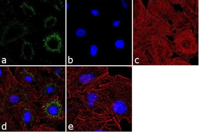
Immunocytochemistry/ Immunofluorescence - Anti-Proteasome 20S C2/HC2 antibody (ab3325)
Immunofluorescence analysis of 70% confluent log phase MDA-MB-231 (Human breast adenocarcinoma cell line) cells labeling Proteasome 20S C2/HC2 (green) with ab3325 at 2 µg/mL. The cells were fixed with 4% paraformaldehyde for 10 minutes, permeabilized with 0.1% Triton™ X-100 for 10 minutes, and blocked with 1% BSA for 1 hour at room temperature. The cells were labeled with ab3325 in 0.1% BSA and incubated for 3 hours at room temperature and then labeled with Goat anti-Rabbit IgG (H+L) secondary antibody, Alexa Fluor® 488 conjugate at 1/2000 dilution for 45 minutes at room temperature (Panel a: green). Nuclei (Panel b: blue) were stained with DAPI. F-actin (Panel c: red) was stained with Alexa Fluor® 555 Rhodamine Phalloidin. Panel d represents the merged image showing cytoplasmic localization. Panel e shows the control without primary antibody. The images were captured at 60X magnification.

Western blot - Anti-Proteasome 20S C2/HC2 antibody (ab3325)
All lanes : Anti-Proteasome 20S C2/HC2 antibody (ab3325) at 2 µg/ml
Lane 1 : Untransfected Hep G2 whole cell extract.
Lane 2 : Proteasome 20S C2/HC2 non-targeting scrambled siRNA transfected Hep G2 whole cell extract.
Lane 3 : Proteasome 20S C2/HC2 knockdown Hep G2 whole cell extract.
Secondary
All lanes : Goat anti-Rabbit IgG (H+L) Superclonal™ Recombinant Secondary Antibody, HRP at 1/4000 dilution
Additional bands at: 45 kDa. We are unsure as to the identity of these extra bands.

Western blot - Anti-Proteasome 20S C2/HC2 antibody (ab3325)
Anti-Proteasome 20S C2/HC2 antibody (ab3325) at 3 µg/ml + CHO (Chinese hamster ovary cell line) whole cell lysate


