Anti-Proteasome 20S LMP7抗体
参阅全部 Proteasome 20S LMP7 一抗
兔多克隆抗体to Proteasome 20S LMP7
Rabbit
适用于: WB, ICCmore details
与反应: Human
预测可用于: Sheep, Cow![]()
Synthetic peptide corresponding to Human Proteasome 20S LMP7 aa 250-350.
Database link: P28062
The Life Science industry has been in the grips of a reproducibility crisis for a number of years. Abcam is leading the way in addressing this with our range of recombinant monoclonal antibodies and knockout edited cell lines for gold-standard validation. Please check that this product meets your needs before purchasing.
If you have any questions, special requirements or concerns, please send us an inquiry and/or contact our Support team ahead of purchase. Recommended alternatives for this product can be found below, along with publications, customer reviews and Q&As
Liquid
Shipped at 4°C. Store at +4°C short term (1-2 weeks). Upon delivery aliquot. Store at -20°C or -80°C. Avoid freeze / thaw cycle.
Constituents: 0.1% BSA, 99% PBS
浓度
100 µg 浓度为 1 mg/ml
Immunogen affinity purified
多克隆
IgG
Abpromise™承诺保证使用ab3329于以下的经测试应用
“应用说明”部分 下显示的仅为推荐的起始稀释度;实际最佳的稀释度/浓度应由使用者检定。
| 应用 | Ab评论 | 说明 |
|---|---|---|
| WB | (12) | Use a concentration of 1 - 3 µg/ml. Detects a band of approximately 20 kDa. |
| ICC | Use a concentration of 5 µg/ml. |
Entrez Gene: 5696 Human
Entrez Gene: 100174905 Sheep
Omim: 177046 Human
SwissProt: P28062 Human
Unigene: 180062 Human
Large multifunctional peptidase 7 antibody
Large multifunctional protease 7 antibody
LMP 7 antibody
LMP7 antibody
Low molecular mass protein 7 antibody
Low molecular weight protein 7 antibody
Macropain subunit C13 antibody
MGC1491 antibody
Multicatalytic endopeptidase complex subunit C13 antibody
NKJO antibody
OTTHUMP00000062981 antibody
Protease component C13 antibody
Proteasome (prosome macropain) subunit beta type 8 antibody
Proteasome (prosome, macropain) subunit, beta type, 8 (large multifunctional peptidase 7) antibody
Proteasome beta 8 subunit antibody
Proteasome catalytic subunit 3i antibody
Proteasome component C13 antibody
Proteasome related gene 7 antibody
Proteasome subunit beta 5i antibody
Proteasome subunit beta 8 antibody
Proteasome subunit beta type 8 antibody
Proteasome subunit beta type antibody
Proteasome subunit beta type-8 antibody
Proteasome subunit beta-5i antibody
Proteasome subunit Y2 antibody
PSB8_HUMAN antibody
PSMB 8 antibody
PSMB5i antibody
PSMB8 antibody
Really interesting new gene 10 protein antibody
RING 10 antibody
RING10 antibody
Y2 antibody
ALDD antibody
D6S216 antibody
D6S216E antibody

Western blot - Anti-Proteasome 20S LMP7 antibody (ab3329)
All lanes : Anti-Proteasome 20S LMP7 antibody (ab3329) at 2 µg/ml
Lane 1 : HeLa whole cell extracts
Lane 2 : HeLa treated with IFN gamma (100ng/ml IFN gamma for 72h) whole cell extracts
Lane 3 : U-937 whole cell extracts
Lane 4 : Raji whole cell extracts
Lane 5 : Ramos whole cell extracts
Lysates/proteins at 30 µg per lane.
Secondary
All lanes : Goat anti-Rabbit IgG (H+L) HRP conjugate at 1/2500 dilution
Observed band size: 20 kDawhy is the actual band size different from the predicted?
Detected by chemiluminescence.

Immunocytochemistry - Anti-Proteasome 20S LMP7 antibody (ab3329)
Immunocytochemistry/ Immunofluorescence analysis of HeLa cells labeling Proteasome 20S LMP7 with ab3329 at 5µg/ml. The cells were fixed with 4% paraformaldehyde for 15 minutes, permeabilized with 0.1% Triton X-100 in TBS for 10 minutes, and blocked with 3% Blocker BSA in PBS for 15 minutes at room temperature. Cells were stained with or without Anti-Proteasome 20S LMP7 antibody (ab3329), at a concentration of 5µg/ml for 1 hour at room temperature, and then incubated with a Alexa Fluor® 488 goat anti-rabbit IgG secondary antibody at a dilution of 1/1000 for 1 hour s at room temperature (both panels, green). Nuclei (both panels, blue) were stained with Hoechst 33342 dye.
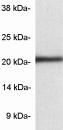
Western blot - Anti-Proteasome 20S LMP7 antibody (ab3329)
Western blot of proteasome 20S LMP7 from HeLa cell extract using ab3329.

Immunocytochemistry - Anti-Proteasome 20S LMP7 antibody (ab3329)
ICC/IF image of ab3329 stained MCF7 cells. The cells were 4% formaldehyde fixed (10 min) and then incubated in 1%BSA / 10% normal goat serum / 0.3M glycine in 0.1% PBS-Tween for 1h to permeabilise the cells and block non-specific protein-protein interactions. The cells were then incubated with the antibody (ab3329, 5µg/ml) overnight at +4°C. The secondary antibody (green) was Alexa Fluor® 488 goat anti-rabbit IgG (H+L) used at a 1/1000 dilution for 1h. Alexa Fluor® 594 WGA was used to label plasma membranes (red) at a 1/200 dilution for 1h. DAPI was used to stain the cell nuclei (blue) at a concentration of 1.43µM.


