| 存储条件 |
|---|
| Product Name | Mouse monoclonal antibody to p23 |
|---|---|
| Antibody Type | Primary Antibodies |
| Immunogen | A synthesized peptide derived from human p23. |
| Clonality | monoclonal |
|---|---|
| Isotype | IgG1 |
| Host Species | Mouse |
| Tested Applications | ICC/IFIHCWB |
| WB:1:500-1:3000 IHC:1:50-1:200 ICC/IF:1:100-1:500 | |
| Species Reactivity | Human |
| Concentration | 1mg/ml |
| Purification | Affinity purified |
| Gene Symbol | PTGES3 |
|---|---|
| Gene Synonyms | P23 TEBP cPGES |
| Gene Full Name | prostaglandin E synthase 3 |
| Gene Summary | This gene encodes an enzyme that converts prostaglandin endoperoxide H2 (PGH2) to prostaglandin E2 (PGE2). This protein functions as a co-chaperone with heat shock protein 90 (HSP90), localizing to response elements in DNA and disrupting transcriptional activation complexes. Alternative splicing results in multiple transcript variants. There are multiple pseudogenes of this gene on several different chromosomes. [provided by RefSeq, Feb 2016] |
| Alternative Names | Co chaperone p23 cPGES Cytosolic prostaglandin E synthase Cytosolic prostaglandin E2 synthase Hsp90 co chaperone Hsp90 co-chaperone P23 Progesterone receptor complex Progesterone receptor complex p23 Prostaglandin E synthase 3 (cytosolic) Prostaglandin E synthase 3 PTGES 3 PTGES3 Sid 3177 TEBP TEBP_HUMAN Telomerase binding protein p23 Telomerase-binding protein p23 Unactive progesterone receptor 23 kD |
| Molecular Weight(MW) | 19kDa |
| Cellular Localization | Cytoplasm. |
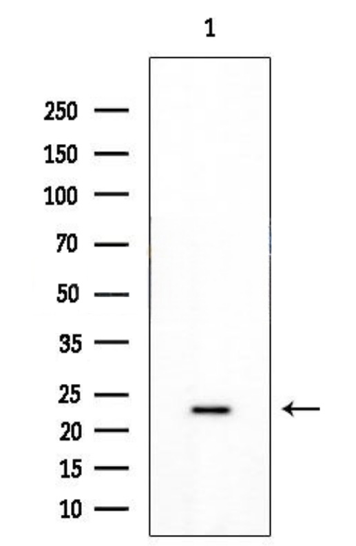
WB
Western blot analysis of extracts from Hela cells, using p23 Mouse Monoclonal Antibody at 1/1000 dilution. Observed bands:23kDa.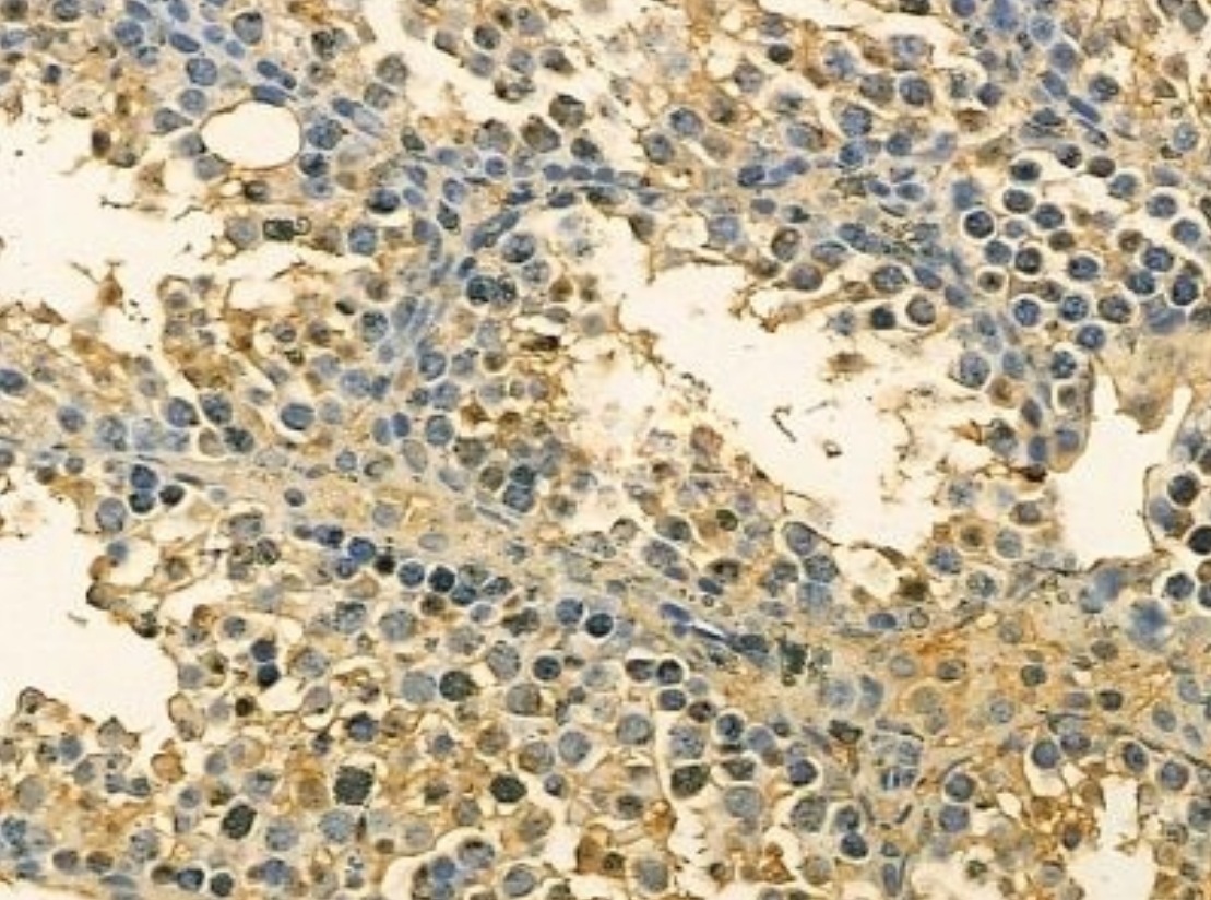
IHC
p23 Mouse Monoclonal Antibody at 1/100 staining mouse testis tissue by IHC-P. The sample was formaldehyde fixed and a heat mediated antigen retrieval step in citrate buffer was performed. The sample was then blocked and incubated with the primary antibody at 4°C overnight. An HRP conjugated anti-mouse antibody was used as the secondary antibody.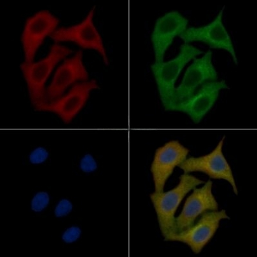
ICC/IF
p23 Mouse Monoclonal Antibody staining Hela cells by IF/ICC. The samples were fixed with PFA and permeabilized in 0.1% Triton X-100, then blocked in 10% serum for 45 minutes at 25°C. Samples were then incubated with primary Ab and rabbit anti-beta tubulin Ab for 1 hour at 37°C. An AlexaFluor594 conjugated goat anti-mouse IgG Ab(Red) and an AlexaFluor488 conjugated goat anti-rabbit IgG Ab(Green) were used as the secondary antibody. The nuclear counter stain is DAPI (blue).| Application Notes | WB:1:500-1:3000 IHC:1:50-1:200 ICC/IF:1:100-1:500 |
|---|
| Form | Liquid |
|---|---|
| Storage Instructions | Store at -20 °C. Stable for 12 months from date of receipt. |
| Storage Buffer | Mouse IgG1 in phosphate buffered saline (without Mg2+ and Ca2+), pH 7.4, 150mM NaCl, 0.02% sodium azide and 50% glycerol. |
抱歉,暂无相关文献
抱歉,暂无浏览记录







