| 存储条件 |
|---|
| Product Name | CD45 Recombinant Rabbit Monoclonal Antibody |
|---|---|
| Antibody Type | Primary Antibodies |
| Immunogen | Recombinant protein within Human CD45 aa 1,210-1,306 / 1,306. |
| Clonality | Monoclonal |
|---|---|
| Isotype | IgG |
| Host Species | Rabbit |
| Tested Applications | FCIF-PIHCWB |
| WB:1:500-1:1000 IHC:1:1000 IF-P:1:500 FC:1:50-1:100 | |
| Species Reactivity | Human |
| Concentration | 1mg/ml |
| Purification | Protein A |
| Gene Symbol | PTPRC |
|---|---|
| Gene Synonyms | LCA LY5 B220 CD45 L-CA T200 CD45R GP180 IMD105 |
| Gene Full Name | protein tyrosine phosphatase receptor type C |
| Gene Summary | The protein encoded by this gene is a member of the protein tyrosine phosphatase (PTP) family. PTPs are known to be signaling molecules that regulate a variety of cellular processes including cell growth, differentiation, mitosis, and oncogenic transformation. This PTP contains an extracellular domain, a single transmembrane segment and two tandem intracytoplasmic catalytic domains, and thus is classified as a receptor type PTP. This PTP has been shown to be an essential regulator of T- and B-cell antigen receptor signaling. It functions through either direct interaction with components of the antigen receptor complexes, or by activating various Src family kinases required for the antigen receptor signaling. This PTP also suppresses JAK kinases, and thus functions as a regulator of cytokine receptor signaling. Alternatively spliced transcripts variants of this gene, which encode distinct isoforms, have been reported. [provided by RefSeq, Jun 2012] |
| Alternative Names | B220 antibody CD 45 antibody CD45 antibody CD45 antigen antibody CD45R antibody GP180 antibody L-CA antibody LCA antibody Leukocyte common antigen antibody loc antibody B220 antibody CD 45 antibody CD45 antibody CD45 antigen antibody CD45R antibody GP180 antibody L-CA antibody LCA antibody Leukocyte common antigen antibody loc antibody Ly-5 antibody LY5 antibody Ly5 homolog of antibody Lyt-4 antibody OTTHUMP00000033813 antibody OTTHUMP00000033816 antibody OTTHUMP00000033817 antibody OTTHUMP00000038574 antibody Protein tyrosine phosphatase receptor type c polypeptide antibody Protein tyrosine phosphatase receptor type C antibody protein tyrosine phosphatase receptor type C antibody Protein tyrosine phosphatase receptor type c polypeptide antibody Ptprc antibody PTPRC_HUMAN antibody Receptor-type tyrosine-protein phosphatase C antibody T200 antibody T200 glycoprotein antibody T200 leukocyte common antigen antibody |
| Molecular Weight(MW) | 147kDa(Observed band size: 200kDa) |
| Cellular Localization | Cell junction, Cell membrane, Membrane |
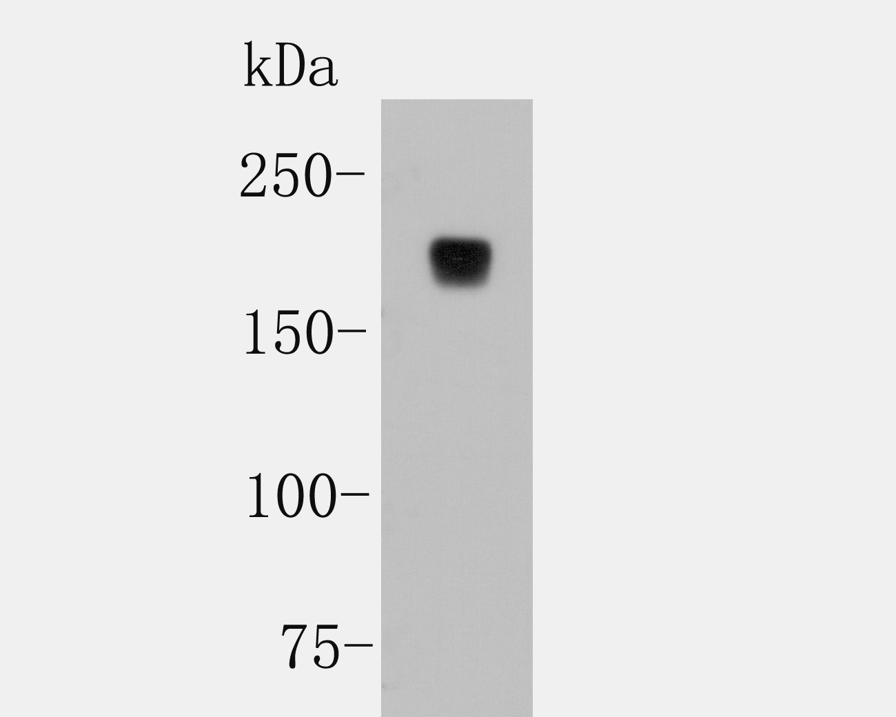
WB
Western blot analysis of CD45 on Jurkat cell lysates with Rabbit anti-CD45 antibody at 1/1000 dilution. Lysates/proteins at 10 µg/Lane. Exposure time: 2 minutes; 8% SDS-PAGE gel. Proteins were transferred to a PVDF membrane and blocked with 5% NFDM/TBST for 1 hour at room temperature. The primary antibody at 1/1000 dilution was used in 5% NFDM/TBST at room temperature for 2 hours. Goat Anti-Rabbit IgG - HRP Secondary Antibody at 1:5,000 dilution was used for 1 hour at room temperature.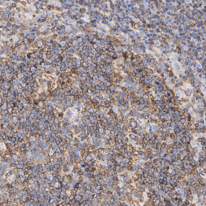
IHC
Immunohistochemical analysis of paraffin-embedded human tonsil tissue with Rabbit anti-CD45 antibody at 1/200 dilution. The section was pre-treated using heat mediated antigen retrieval with Tris-EDTA buffer (pH 9.0) for 20 minutes. The tissues were blocked in 1% BSA for 20 minutes at room temperature, washed with ddH2O and PBS, and then probed with the primary antibody at 1/200 dilution for 1 hour at room temperature. The detection was performed using an HRP conjugated compact polymer system. DAB was used as the chromogen. Tissues were counterstained with hematoxylin and mounted with DPX.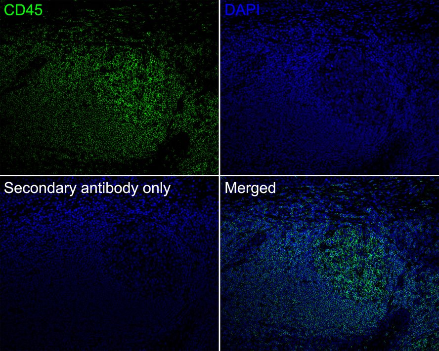
IF-P
Immunofluorescence analysis of paraffin-embedded human tonsil tissue labeling CD45 with Rabbit anti-CD45 antibody at 1/500 dilution. The section was pre-treated using heat mediated antigen retrieval with Tris-EDTA buffer (pH 9.0) for 20 minutes. The tissues were blocked in 10% negative goat serum for 1 hour at room temperature, washed with PBS, and then probed with the primary antibody (green) at 1/500 dilution overnight at 4 ℃, washed with PBS. Goat Anti-Rabbit IgG H&L (iFluor™ 488) was used as the secondary antibody at 1/1,000 dilution. Nuclei were counterstained with DAPI (blue).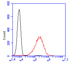
FC
Flow cytometric analysis of CD45 was done on Jurkat cells. The cells were fixed, permeabilized and stained with the primary antibody (1/100) (red). After incubation of the primary antibody at room temperature for an hour, the cells were stained with a Alexa Fluor 488-conjugated goat anti-rabbit IgG Secondary antibody at 1/500 dilution for 30 minutes.Unlabelled sample was used as a control (cells without incubation with primary antibody; black).| Application Notes | WB:1:500-1:1000 IHC:1:1000 IF-P:1:500 FC:1:50-1:100 |
|---|
| Form | Liquid |
|---|---|
| Storage Instructions | Store at +4℃ after thawing. Aliquot store at -20℃. Avoid repeated freeze / thaw cycles. |
| Storage Buffer | 1*TBS (pH7.4), 0.05% BSA, 40% Glycerol. Preservative: 0.05% Sodium Azide. |
抱歉,暂无相关文献







