| 存储条件 |
|---|
| Product Name | Caspase-8 (p18) Recombinant Rabbit Monoclonal Antibody |
|---|---|
| Antibody Type | Primary Antibodies |
| Immunogen | Synthetic peptide corresponding to residues surrounding Asp387 of mouse caspase-8 protein. |
| Clonality | Monoclonal |
|---|---|
| Isotype | IgG |
| Host Species | Rabbit |
| Tested Applications | ICC/IFWB |
| WB:1:2000 ICC:1:100 | |
| Species Reactivity | Mouse |
| Concentration | 1mg/ml |
| Purification | Protein A |
| Gene Symbol | Casp8 |
|---|---|
| Gene Synonyms | MACH Mch5 FLICE CASP-8 |
| Gene Full Name | caspase 8 |
| Gene Summary | This gene is part of a family of caspases, aspartate-specific cysteine proteases well studied for their involvement in immune and apoptosis signaling. This protein, an initiator of apoptotic cell death, is activated by death-inducing tumor necrosis family receptors and targets downstream effectors. In mouse deficiency of this gene can cause embryonic lethality. This protein may have a role in embryogenesis. Alternative splicing results in multiple transcript variants that encode different protein isoforms. [provided by RefSeq, Apr 2013] |
| Alternative Names | ALPS2B antibody Amyotrophic lateral sclerosis 2 chromosomal region candidate gene 12 protein antibody Apoptosis related cysteine peptidase antibody Apoptotic cysteine protease antibody Apoptotic protease Mch-5 antibody Apoptotic protease Mch5 antibody CAP 4 antibody CAP4 antibody CASP-8 antibody CASP8 antibody ALPS2B antibody Amyotrophic lateral sclerosis 2 chromosomal region candidate gene 12 protein antibody Apoptosis related cysteine peptidase antibody Apoptotic cysteine protease antibody Apoptotic protease Mch-5 antibody Apoptotic protease Mch5 antibody CAP 4 antibody CAP4 antibody CASP-8 antibody CASP8 antibody CASP8_HUMAN antibody Caspase 8 antibody Caspase 8 apoptosis related cysteine peptidase antibody Caspase IIX antibody Caspase-8 subunit p10 antibody caspase8 antibody CED 3 antibody FADD Homologous ICE/CED3 Like Protease antibody FADD Like ICE antibody FADD-homologous ICE/CED-3-like protease antibody FADD-like ICE antibody FLICE antibody FLJ17672 antibody ICE-like apoptotic protease 5 antibody MACH alpha 1/2/3 protein antibody MACH antibody MACH beta 1/2/3/4 protein antibody MACH5 antibody MCH 5 antibody MCH5 antibody MGC78473 antibody MORT1 associated ced 3 homolog antibody MORT1 associated CED3 homolog antibody MORT1-associated CED-3 homolog antibody OTTHUMP00000163717 antibody OTTHUMP00000163720 antibody OTTHUMP00000163724 antibody OTTHUMP00000163725 antibody OTTHUMP00000165062 antibody OTTHUMP00000165063 antibody OTTHUMP00000165064 antibody OTTHUMP00000206552 antibody OTTHUMP00000206582 antibody |
| Molecular Weight(MW) | 43/18kDa |
| Cellular Localization | Cytoplasm. Nucleus. |
WB
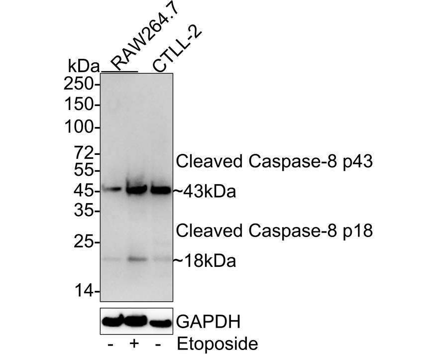
Western blot analysis of Caspase-8 (p18) on different lysates with Rabbit anti-Caspase-8 (p18) antibody at 1/2,000 dilution. Lane 1: RAW264.7 cell lysate, Lane 2: RAW264.7 treated with 1μM Etoposide for 18 hours cell lysate, Lane 3: CTLL-2 cell lysate. Lysates/proteins at 30 µg/Lane. Exposure time: 1 minute 20 seconds; 4-20% SDS-PAGE gel. Proteins were transferred to a PVDF membrane and blocked with 5% NFDM/TBST for 1 hour at room temperature. The primary antibody at 1/2,000 dilution was used in 5% NFDM/TBST at 4℃ overnight. Goat Anti-Rabbit IgG - HRP Secondary Antibody at 1/50,000 dilution was used for 1 hour at room temperature.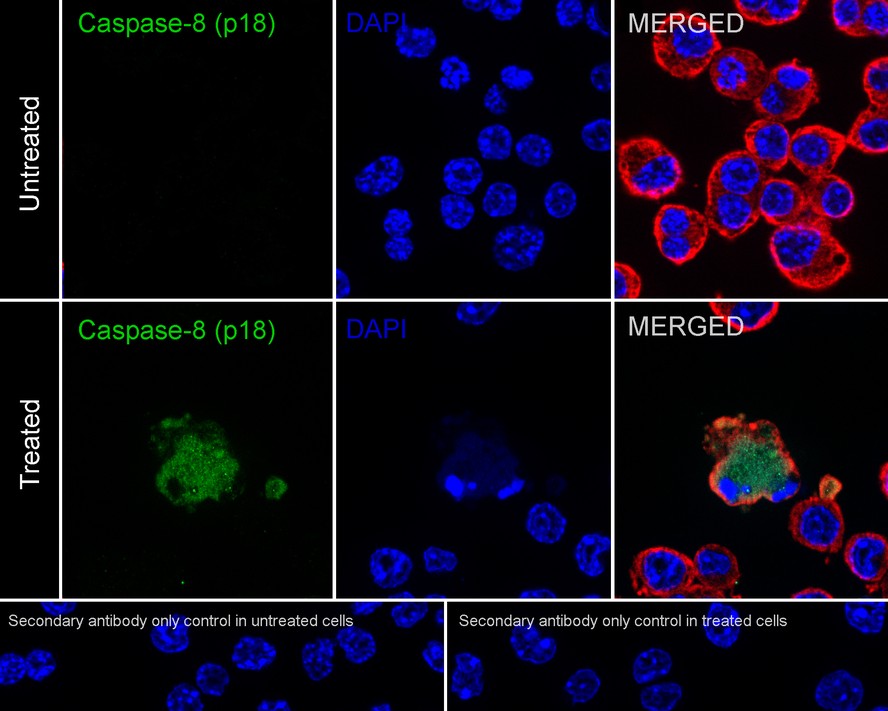
ICC/IF
Immunocytochemistry analysis of RAW264.7 cells treated with 1μM Etoposide for 18 hours labeling Caspase-8 (p18) with Rabbit anti-Caspase-8 (p18) antibody at 1/100 dilution. Cells were fixed in 4% paraformaldehyde for 20 minutes at room temperature, permeabilized with 0.1% Triton X-100 in PBS for 5 minutes at room temperature, then blocked with 1% BSA in 10% negative goat serum for 1 hour at room temperature. Cells were then incubated with Rabbit anti-Caspase-8 (p18) antibody at 1/100 dilution in 1% BSA in PBST overnight at 4 ℃. Goat Anti-Rabbit IgG H&L (iFluor™ 488) was used as the secondary antibody at 1/1,000 dilution. PBS instead of the primary antibody was used as the secondary antibody only control. Nuclear DNA was labelled in blue with DAPI. Beta tubulin (red) was stained at 1/100 dilution overnight at +4℃. Goat Anti-Mouse IgG H&L (iFluor™ 594) was used as the secondary antibody at 1/1,000 dilution.| Application Notes | WB:1:2000 ICC:1:100 |
|---|
| Form | Liquid |
|---|---|
| Storage Instructions | Store at +4℃ after thawing. Aliquot store at -20℃. Avoid repeated freeze / thaw cycles. |
| Storage Buffer | PBS (pH7.4), 0.1% BSA, 40% Glycerol. Preservative: 0.05% Sodium Azide. |
抱歉,暂无相关文献







