| 存储条件 |
|---|
| Product Name | Rabbit polyclonal antibody to Phospho-LAT (Tyr191)[Tyr220] |
|---|---|
| Antibody Type | Primary Antibodies |
| Immunogen | A synthesized peptide derived from human LAT(Accession O43561), corresponding to amino acid residues around phosphorylated Tyr220. |
| Modification | p-Tyr191 |
| Modification Notes | Phospho-LAT (Tyr191) Antibody detects endogenous levels of LAT only when phosphorylated at Tyr220, which site historically referenced as Tyr191. |
| Clonality | Polyclonal |
|---|---|
| Isotype | IgG |
| Host Species | Rabbit |
| Tested Applications | ICC/IFIHCWB |
| WB:1:500-1:2000 IHC:1:50-1:200 ICC:1:100-1:500 | |
| Species Reactivity | HumanMouse |
| Concentration | 1mg/ml |
| Purification | Affinity purified |
| Gene Symbol | LAT |
|---|---|
| Gene Synonyms | LAT1 pp36 IMD52 |
| Gene Full Name | linker for activation of T cells |
| Gene Summary | The protein encoded by this gene is phosphorylated by ZAP-70/Syk protein tyrosine kinases following activation of the T-cell antigen receptor (TCR) signal transduction pathway. This transmembrane protein localizes to lipid rafts and acts as a docking site for SH2 domain-containing proteins. Upon phosphorylation, this protein recruits multiple adaptor proteins and downstream signaling molecules into multimolecular signaling complexes located near the site of TCR engagement. Alternative splicing results in multiple transcript variants encoding different isoforms. [provided by RefSeq, Jul 2008] |
| Alternative Names | 36 kDa phospho tyrosine adapter protein 36 kDa phospho-tyrosine adapter protein 36 kDa phospho-tyrosine adaptor protein LAT 1 lat LAT_HUMAN LAT1 Linker for activation of T cells Linker for activation of T cells family member 1 linker for activation of T cells transmembrane adaptor Linker for activation of T-cells family member 1 p36 38 p36-38 pp36 |
| Molecular Weight(MW) | 28kDa |
| Cellular Localization | Cell membrane |
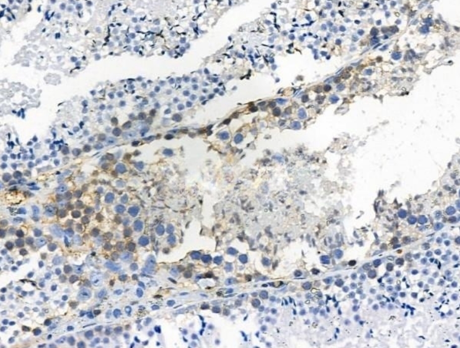
IHC
Phospho-LAT (Tyr191)[Tyr220] antibody at 1/100 staining Mouse testis tissue by IHC-P. The sample was formaldehyde fixed and a heat mediated antigen retrieval step in citrate buffer was performed. The sample was then blocked and incubated with the primary antibody at 4°C overnight. An HRP conjugated anti-Rabbit antibody was used as the secondary antibody.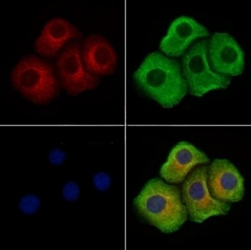
ICC/IF
Phospho-LAT (Tyr191)[Tyr220] antibody staining Hela cells(4h of LPS treatment) by IF/ICC. The samples were fixed with PFA and permeabilized in 0.1% Triton X-100,then blocked in 10% serum for 45 minutes at 25°C. Samples were then incubated with primary Ab(1:200) and mouse anti-beta tubulin Ab(1:200) for 1 hour at 37°C. An AlexaFluor594 conjugated goat anti-rabbit IgG(H+L) Ab(Red) and an AlexaFluor488 conjugated goat anti-mouse IgG(H+L) Ab(Green) were used as the secondary antibody. The nuclear counter stain is DAPI(blue).| Application Notes | WB:1:500-1:2000 IHC:1:50-1:200 ICC:1:100-1:500 |
|---|
| Form | Liquid |
|---|---|
| Storage Instructions | Store at -20 °C. Stable for 12 months from date of receipt. |
| Storage Buffer | Rabbit IgG in phosphate buffered saline , pH 7.4, 150mM NaCl, 0.02% sodium azide and 50% glycerol. |
抱歉,暂无相关文献







