Anti-GFP抗体
参阅全部 GFP 一抗
山羊多克隆抗体to GFP
Goat
No reaction was observed against Human, Mouse or Rat serum proteins.
适用于: WB, IP, ELISA, ICC/IF, IHC-P, IHC-FrFl, IHC-Frmore details
与反应: Species independent
Recombinant full length protein corresponding to Aequorea victoria GFP aa 1-250.
Database link: P42212
IHC: E5.5 Hex-GFP transgenic mouse embryo. WB: Pure GFP protein, or cells known to overexpress GFP.
This anti-GFP antibody cross reacts with eGFP .
The Life Science industry has been in the grips of a reproducibility crisis for a number of years. Abcam is leading the way in addressing this with our range of recombinant monoclonal antibodies and knockout edited cell lines for gold-standard validation. Please check that this product meets your needs before purchasing.
If you have any questions, special requirements or concerns, please send us an inquiry and/or contact our Support team ahead of purchase. Recommended alternatives for this product can be found below, along with publications, customer reviews and Q&As
Liquid
Shipped at 4°C. Store at +4°C short term (1-2 weeks). Upon delivery aliquot. Store at -20°C. Avoid freeze / thaw cycle.
pH: 7.2
Preservative: 0.01% Sodium azide
Constituents: 0.424% Potassium phosphate solution, 0.88% Sodium chloride
浓度
批次浓度范围 100 µg 浓度为 1 - 1.1 mg/ml
Affinity purified
GFP antibody was prepared from monospecific antiserum by immunoaffinity chromatography using Green Fluorescent Protein (Aequorea victoria) coupled to agarose beads followed by solid phase adsorption(s) to remove any unwanted reactivities.
多克隆
IgG
Abpromise™承诺保证使用ab6673于以下的经测试应用
“应用说明”部分 下显示的仅为推荐的起始稀释度;实际最佳的稀释度/浓度应由使用者检定。
| 应用 | Ab评论 | 说明 |
|---|---|---|
| WB | (3) | 1/1000 - 1/10000. (for immunoprecipitated GFP, see Abreview). |
| IP | Use at an assay dependent concentration. | |
| ELISA | 1/10000 - 1/30000. This antibody can be used to detect GFP by ELISA (sandwich or capture) for the direct binding of antigen and recognizes wild type, recombinant and enhanced forms of GFP. | |
| ICC/IF | (1) | 1/500. |
| IHC-P | (10) | 1/200 - 1/1000. |
| IHC-FrFl | Use at an assay dependent concentration. | |
| IHC-Fr | (6) | 1/200 - 1/1000. |
| IF | Use at an assay dependent concentration. |
GFP antibody
Green fluorescent protein antibody

IHC - Wholemount - Anti-GFP antibody (ab6673)
Immunofluorescence Microscopy using ab6673.
Tissue: Drosophila melanogaster late stage embryonic central nervous system.
Fixation: 0.5% PFA.
Antigen retrieval: Not required.
Primary antibody: Anti-GFP antibody at a 1/1,000 for 1 h at RT.
Secondary antibody: AlexaFluor 488™ conjugated anti-Goat antibody at 1/300 for 45 minutes at RT.
Panel A: shows a lateral view (ventral left).
Panels B and C: shows ventral views of whole mount embryos at 63x magnification (plus 2x digital zoom).
In all panels, anterior is up.
Staining: tau-GFP cell bodies (large arrowhead) and axons of motorneurons (arrow) and interneurons (small arrowhead) as green fluorescent signal.

Immunohistochemistry - Free Floating - Anti-GFP antibody (ab6673)Suarez-Bregua et al PLoS One. 2017 Oct 17;12(10):e0186444. doi: 10.1371/journal.pone.0186444. eCollection 2017. Fig 1. Reproduced under the Creative Commons license http://creativecommons.org/licenses/by/4.0/
Pth4:eGFP transgenic zebrafish embryos at 1 and 2 dpf were fixed with 4% PFA and washed in PBST. They were then washed in PBDT (1% BSA, 1% DMSO, 0.1% Triton X-100 in PBS, pH 7.4), blocked in 10% normal goat serum/PBDT, and incubated overnight at 4°C with primary antibodies to HuC/D (1/100) and GFP (1/400, Abcam ab6673). Further PBST washes and blocking were followed by secondary antibodies overnight at 4°C. Hoechst 34580 was added to stain nuclei (1/2500). After further PBDT and PBS washes, embryos were mounted for confocal imaging.
Abbreviation: e, eye; hy, hypothalamus; m, midbrain; sc, spinal cord. Scale bars: 100 μm (A-C) 50 μm (D-G).

Immunocytochemistry/ Immunofluorescence - Anti-GFP antibody (ab6673)Borkowska et al PLoS One. 2016 May 31;11(5):e0156082. doi: 10.1371/journal.pone.0156082. eCollection 2016. Fig 5. Reproduced under the Creative Commons license http://creativecommons.org/licenses/by/4.0/
In utero electroporation of Disc1 and Disc1-100P constructs into wild-type neocortex and analysis at P21.
(Panels D-E”) Expression of the constructs was assessed.
(Panels D-D'') 2 days after transfection in vitro.
(Panels E-E'') at P21 in vivo.
Immunochemistry for FLAG and GFP showed that constructs encoding either WT Disc1, the Disc1-100P variant, or GFP alone, expressed these protein species in transfected HEK-293 cells in vitro (Fig 5D–5D”) and in P21 postmitotic cortical neurons in vivo (Fig 5E–5E”)

Immunohistochemistry (Frozen sections) - Anti-GFP antibody (ab6673)Goldman et al PLoS One. 2018 Jan 12;13(1):e0191245. doi: 10.1371/journal.pone.0191245. eCollection 2018. Fig 5. Reproduced under the Creative Commons license https://creativecommons.org/publicdomain/zero/1.0/
Immunofluorescence for assessment of GFP+ myofibers in rat tissue.
VML affected muscle from the 50% MG + HA+LMN group were probed for the presence of GFP. GFP+ fibers were detected in a qualitatively similar magnitude at both 2 and 8 weeks post-injury indicating viable engraftment of donor derived muscle progenitor cells. Scale bars are 1mm for whole mount images, 50 μm for regions of interest.
A portion of the TA muscle from the defect region was embedded in a talcum-based gel, frozen in 2-methylbutane, and supercooled in liquid nitrogen. Cryosections (8 μm) were prepared and stained using standard protocols for hematoxylin & eosin.
ab6673 used at a 1/100 dilution.

Immunohistochemistry (Formalin/PFA-fixed paraffin-embedded sections) - Anti-GFP antibody (ab6673)Cedeno et al PLoS One. 2017 Sep 21;12(9):e0185196. doi: 10.1371/journal.pone.0185196. eCollection 2017. Fig 3. Reproduced under the Creative Commons license http://creativecommons.org/licenses/by/4.0/
Mouse small intestines were washed with DPBS and fixed overnight at 4°C in Zinc formalin. Following sectioning and tissue deparaffanization, antigen retrieval was performed with 10mM Tris base (pH 9.0) buffer using a pressure cooker.
For immunohistochemistry, sections were quenched of endogenous peroxidases by 3% H2O2, and sequentially blocked with Avidin D, biotin, and protein blocking reagents. Primary antibody incubation was conducted at 4°C overnight. Secondary biotinylated antibody was added at a dilution of 1/200, and incubated 2 hours at room temperature. Finally, sections were stained according to the ABC peroxidase protocol and counterstained with hematoxylin.
ab6673 used at a 1/200 dilution.
Panel D: Representative anti-eGFP immunofluorescence of macroH2A WT and DKO jejunum counterstained with DAPI (blue).

Western blot - Anti-GFP antibody (ab6673)
All lanes : Anti-GFP antibody (ab6673) at 1 µg/ml (o/n at 4degC)
Lane 1 : HEK-293 (Human epithelial cell line from embryonic kidney) lysate at 10 µg
Lane 2 : HeLa (Human epithelial cell line from cervix adenocarcinoma) lysate at 10 µg
Lane 3 : CHO/K1 lysate at 10 µg
Lane 4 : MDA-MB-231 (Human breast adenocarcinoma cell line) lysate at 10 µg
Lane 5 : A431 (Human epidermoid carcinoma cell line) lysate at 10 µg
Lane 6 : Jurkat (Human T cell leukemia cell line from peripheral blood) lysate at 10 µg
Lane 7 : NIH/3T3 (Mouse embryo fibroblast cell line) lysate
Lane 8 : E-coli HCP control, 50 ng
Lane 9 : FLAG Positive control lysate at 10 µg
Lane 10 : Red fluorescent protein, 50 ng
Lane 11 : Green fluorescent protein, 50 ng
Lane 12 : Glutathinoe-S-Transferase protein, 50 ng
Lane 13 : Maltose Binding protein, 50 ng
Secondary
All lanes : Peroxidase goat secondary antibody, 60 min at RT at 1/30000 dilution
Blocking Buffer: 1% Casein-TTBS for 30 min at RT.

Immunofluorescence - Anti-GFP antibody (ab6673)
E5.5 Hex-GFP transgenic mouse embryo stained for GFP using ab6673 at 1/500 dilution. Secondary antibody is a fluorochrome conjugated anti-goat IgG secondary antibody at 1/10,000 for 45 min at RT.
Staining: GFP as green fluorescent signal with DAPI blue counterstain.
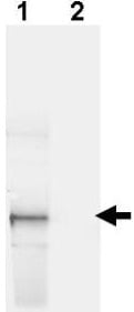
Western blot - Anti-GFP antibody (ab6673)
All lanes : Anti-GFP antibody (ab6673) at 1 µg/ml
Lane 1 : HeLa (Human epithelial cell line from cervix adenocarcinoma) cells
Lane 2 : Mock transfected HeLa cell lysate
Lysates/proteins at 35 µg per lane.
Secondary
All lanes : IRDye® 800 conjugated Donkey-a-Goat IgG [H&L] at 1/2500 dilution
Additional bands at: 33 kDa. We are unsure as to the identity of these extra bands.

Western blot - Anti-GFP antibody (ab6673)This image is courtesy of an anonymous abreview.
All lanes : Anti-GFP antibody (ab6673) at 1/1000 dilution
Lane 1 : MRC5VA lung fibroblast whole cell lysate overexpressing EGFP alone
Lanes 2-3 : MRC5VA lung fibroblast whole cell lysate overexpressing an EGFP fusion protein
Lysates/proteins at 15 µg per lane.
Secondary
All lanes : HRP-conjugated anti-goat polyclonal at 1/10000 dilution
Developed using the ECL technique.
Performed under reducing conditions.
Observed band size: 27,55 kDawhy is the actual band size different from the predicted?
Exposure time: 5 seconds

Immunohistochemistry (Formalin/PFA-fixed paraffin-embedded sections) - Anti-GFP antibody (ab6673)This image is courtesy of Bart Rountree
Immunofluorescence of TGN mouse liver labeling GFP on hepatocytes with ab6673.

Immunohistochemistry (Formalin/PFA-fixed paraffin-embedded sections) - Anti-GFP antibody (ab6673)This image is courtesy of Jeff Klein
Immunohistochemistry of GFP transgenic mouse liver labeling GFP with ab6673.


