Anti-IL-6抗体
参阅全部 IL-6 一抗
兔多克隆抗体to IL-6
Rabbit
ab6672 detects a band at 25 kDa in human lung tissue lysate and mouse spleen tissue lysate, however the signal in mouse tissue is significantly lower. It also binds strongly to a protein at ~55 kDa in human lung tissue extracts, which we believe represents a glycosylated form of IL6. ab6672 also detects several bands in human lung tissue lysate within the region of 30-40 kDa. These may represent heteromers of IL6. Please be aware that this product has low homology with the mouse and rat sequence of IL6 (Rat, 40%; Mouse 41%, UniProt blast) and we therefore cannot guarantee reactivity in these species.
适用于: WB, IHC-Pmore details
与反应: Human
预测可用于: Pig![]()
Recombinant full length protein corresponding to Human IL-6. Produced in E.coli.
Database link: P05231
WB: Recombinant Human IL6 protein (ab101044), lysate of 2 x 10<6 endotoxin-stimulated human peripheral blood mononuclear cells (PBMC)(PBMC are stimulated for 24 hours with 1% (v/v) human serum plus 10 ng/mL E.coli LPS).
IL-6 synonyms: plasmacytoma growth factor (PCT-GF),interferon-a-2 (IFN-a2), monocyte derived human B cellgrowth factor, B cell stimulating factor (BSF-2),hepatocyte stimulating factor (HSF), and interleukinhybridoma/plasmacytoma-1 (IL-HP1).
The Life Science industry has been in the grips of a reproducibility crisis for a number of years. Abcam is leading the way in addressing this with our range of recombinant monoclonal antibodies and knockout edited cell lines for gold-standard validation. Please check that this product meets your needs before purchasing.
If you have any questions, special requirements or concerns, please send us an inquiry and/or contact our Support team ahead of purchase. Recommended alternatives for this product can be found below, along with publications, customer reviews and Q&As
Liquid
Shipped at 4°C. Store at +4°C short term (1-2 weeks). Upon delivery aliquot. Store at -20°C or -80°C. Avoid freeze / thaw cycle.
pH: 7.20
Constituents: 0.424% Potassium phosphate solution, 0.88% Sodium chloride
Whole antiserum
Anti-IL-6 antiserum detects recombinant and native IL-6 present in body fluids and cell supernatants in various assays (ie. IL-1 stimulated IL-6 production from fibroblasts).
多克隆
IgG
Abpromise™承诺保证使用ab6672于以下的经测试应用
“应用说明”部分 下显示的仅为推荐的起始稀释度;实际最佳的稀释度/浓度应由使用者检定。
| 应用 | Ab评论 | 说明 |
|---|---|---|
| WB | (14) | 1/500 - 1/2000. |
| IHC-P | (15) | 1/400 - 1/800. |
Entrez Gene: 3569 Human
Omim: 147620 Human
SwissProt: P05231 Human
Unigene: 654458 Human
B-cell stimulatory factor 2 antibody
BSF 2 antibody
BSF-2 antibody
BSF2 antibody
CDF antibody
CTL differentiation factor antibody
Cytotoxic T cell differentiation factor antibody
Hepatocyte stimulating factor antibody
Hepatocyte stimulatory factor antibody
HGF antibody
HSF antibody
Hybridoma growth factor antibody
Hybridoma growth factor Interferon beta-2 antibody
Hybridoma plasmacytoma growth factor antibody
IFN-beta-2 antibody
IFNB2 antibody
IL 6 antibody
IL-6 antibody
IL6 antibody
IL6_HUMAN antibody
Interferon beta 2 antibody
Interferon beta-2 antibody
Interleukin 6 antibody
Interleukin 6 (interferon beta 2) antibody
Interleukin BSF 2 antibody
Interleukin-6 antibody
Interleukin BSF 2 antibody
B cell differentiation factor antibody
B cell stimulatory factor 2 antibody

Immunohistochemistry (Formalin/PFA-fixed paraffin-embedded sections) - Anti-IL-6 antibody (ab6672)
IHC image of IL6 staining in human lung formalin fixed paraffin embedded tissue section*, performed on a Leica Bond™ system using the standard protocol F. The section was pre-treated using heat mediated antigen retrieval with sodium citrate buffer (pH6, epitope retrieval solution 1) for 20 mins. The section was then incubated with ab6672, 1/400, for 15 mins at room temperature and detected using an HRP conjugated compact polymer system. DAB was used as the chromogen. The section was then counterstained with haematoxylin and mounted with DPX.
For other IHC staining systems (automated and non-automated) customers should optimize variable parameters such as antigen retrieval conditions, primary antibody concentration and antibody incubation times.
*Tissue obtained from the Human Research Tissue Bank, supported by the NIHR Cambridge Biomedical Research Centre

Western blot - Anti-IL-6 antibody (ab6672)
All lanes : Anti-IL-6 antibody (ab6672) at 1/500 dilution
Lane 1 : Human spleen tissue lysate
Lane 2 : Human lung tissue lysate
Lane 3 : Mouse spleen tissue lysate
Lane 4 : Mouse lung tissue lysate
Lysates/proteins at 20 µg per lane.
Performed under reducing conditions.
Additional bands at: 25 kDa, 55 kDa (possible glycosylated form). We are unsure as to the identity of these extra bands.
Tissue lysates were denatured for 10-15 minutes at 90ºC. ab6672 was incubated overnight at 4ºC and the secondary antibody for 1 hour at RT.
ab6672 detects a band at 25 kDa in Human lung tissue lysate and Mouse spleen tissue lysate, however the signal in mouse tissue is significantly lower. It also binds strongly to a protein at ~55 kDa in Human lung tissue extracts, which we believe represents a glycosylated form of IL6. ab6672 also detects several bands in Human lung tissue lysate within the region of 30-40 kDa. These may represent heteromers of IL6.

Immunohistochemistry (Formalin/PFA-fixed paraffin-embedded sections) - Anti-IL-6 antibody (ab6672)Lee EJ et al. Radiation Inhibits Interleukin-12 Production via Inhibition of C-Rel through the Interleukin-6/ Signal Transducer and Activator of Transcription 3 Signaling Pathway in Dendritic Cells. PLoS One 11:e0146463 (2016).
IL6 (Red) and IL-12 (Green) were measured at 1, 3, and 7 days after 10 Gy irradiation of HCa-1 tumors to determine whether irradiation regulates IL-12 and IL6 expression in tumours. ab6672 was used to stain IL6 at 1/100 dilution in immunohistochemical analysis.
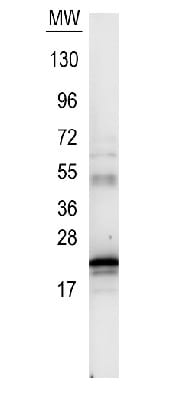
Western blot - Anti-IL-6 antibody (ab6672)
Anti-IL-6 antibody (ab6672) at 1/500 dilution + recombinant human IL-6
Secondary
conjugated anti-Rabbit IgG at 1/40000 dilution
Developed using the ECL technique.
Observed band size: 21 kDawhy is the actual band size different from the predicted?
4-20% Tris-Glycine gel.
The membrane was blocked for 30 minutes with 1% BSA-TBST.


