生物素Anti-GFP抗体
参阅全部 GFP 一抗
生物素山羊多克隆抗体to GFP
Goat
Biotin
Antibody recognizes wild type, recombinant and enhanced forms of GFP (EGFP). No reaction was observed against Human, Mouse and Rat Serum Proteins.
适用于: WB, IP, ICC/IF, Sandwich ELISA, IHC-P, IHC-Frmore details
与反应: Species independent
Recombinant full length protein corresponding to GFP aa 1-246.
Sequence:
MSKGEELFTGVVPILVELDGDVNGHKFSVSGEGEGDATYGKLTLKFICTT GKLPVPWPTL VTTFSYGVQCFSRYPDHMKQHDFFKSAMPEGYVQERTI FFKDDGNYKTRAEVKFEGDTLV NRIELKGIDFKEDGNILGHKLEYNYN SHNVYIMADKQKNGIKVNFKIRHNIEDGSVQLAD HYQQNTPIGDGPVL LPDNHYLSTQSALSKDPNEKRDHMVLLEFVTAAGITHGMDELYK
Database link: P42212
Designed to detect GFP and its variants in immunoblotting and immunoprecipitation.
Biotinamidocaproate N-Hydroxysuccinimide Ester (BAC) Biotin/Protein Ratio: 10-20 BAC molecules per goat IgG molecule.
The Life Science industry has been in the grips of a reproducibility crisis for a number of years. Abcam is leading the way in addressing this with our range of recombinant monoclonal antibodies and knockout edited cell lines for gold-standard validation. Please check that this product meets your needs before purchasing.
If you have any questions, special requirements or concerns, please send us an inquiry and/or contact our Support team ahead of purchase. Recommended alternatives for this product can be found below, along with publications, customer reviews and Q&As
Liquid
Shipped at 4°C. Store at +4°C short term (1-2 weeks). Upon delivery aliquot. Store at -20°C. Avoid freeze / thaw cycle.
Preservative: 0.01% Sodium azide
Constituents: 1% BSA, 0.424% Potassium phosphate solution, 0.88% Sodium chloride
10 mg/mL BSA, immunoglobulin and protease free
浓度
100 µg 浓度为 1 mg/ml
Affinity purified
Anti-GFP was prepared from monospecific antiserum by immunoaffinity chromatography using Green Fluorescent Protein (Aequorea victoria) coupled to agarose beads followed by solid phase adsorption(s) to remove any unwanted reactivities.
Designed to detect GFP and its variants in ELISA (sandwich or capture), immunoblotting and immunoprecipitation.
多克隆
IgG
Abpromise™承诺保证使用ab6658于以下的经测试应用
“应用说明”部分 下显示的仅为推荐的起始稀释度;实际最佳的稀释度/浓度应由使用者检定。
| 应用 | Ab评论 | 说明 |
|---|---|---|
| WB | (2) | 1/2000 - 1/10000. |
| IP | (1) | Use at an assay dependent concentration. |
| ICC/IF | (1) | 1/1000 - 1/5000. |
| Sandwich ELISA | Use at an assay dependent concentration. | |
| IHC-P | (2) | 1/1000 - 1/5000. |
| IHC-Fr | 1/5000. |
GFP antibody
Green fluorescent protein antibody

Western blot - Biotin Anti-GFP antibody (ab6658)
Biotin Anti-GFP antibody (ab6658)
Additional bands at: 28 kDa. We are unsure as to the identity of these extra bands.

Immunohistochemistry (Formalin/PFA-fixed paraffin-embedded sections) - Biotin Anti-GFP antibody (ab6658)This image is a courtesy of Hongwei Shao
ab6658 staining GFP in human melanoma cells recovered from nude mice by Immunocytochemistry/ immunoflurescence. Cells were fixed with formaldehyde, permeabilized with 0.25% Triton X-100 RT for 10min and blocking with commercially available blocking buffer was performed for 30 minutes at RT. Samples were incubated with primary antibody (1:50) for 18 hours at 4°C. An Alexa Fluor®488-conjugated donkey polyclonal to goat IgG was used as secondary antibody at 1/100 dilution. Green color indicates GFP/Fibrolast cells, while red color indicates Ki67 positive cells, most of them are tumor cells (Abcam`s ab15580 was used for the detection).
IHC - Wholemount - Biotin Anti-GFP antibody (ab6658)
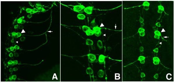
Immunofluorescence Microscopy using ab6658. Tissue: Drosophila melanogaster late stage embryonic central nervous system. Fixation: 0.5% PFA. Antigen retrieval: not required. Primary antibody: Anti-GFP antibody at a 1/1,000 for 1 h at RT. Secondary antibody: AlexaFluor 488™ conjugated anti-Goat antibody at 1/300 for 45 min at RT. Panel A: shows a lateral view (ventral left). Panels B and C: shows ventral views of whole mount embryos at 63x magnification (plus 2x digital zoom). In all panels, anterior is up. Staining: tau-GFP cell bodies (large arrowhead) and axons of motorneurons (arrow) and interneurons (small arrowhead) as green fluorescent signal.

Immunohistochemistry (Frozen sections) - Biotin Anti-GFP antibody (ab6658)
Immunofluorescence Microscopy using ab6658. Tissue: Sf-1:Cre mice crossed to the Z/EG reporter line. Mouse brain (coronal view, 20X magnification). Fixation: 4%PFA/PBS with o/n fixation, and subsequently transferred to a 30% sucrose solution. Antigen retrieval: frozen in OCT freezing medium (Sakura) and cryostat sectioned at 40 microns. Primary antibody: Goat anti- GFP was used at 1/500 dilution in free floating imunnohistochemistry to detect GFP. Secondary antibody: Fluorchrome conjugated Anti-goat IgG secondary antibody was used for detection at 1:10,000 for 45 min at RT.Localization: Sf-1+ neurons and their processes of the ventromedial nucleus of the hypothalamus. Staining: eGFP as green fluorescent signal and sections were counterstained with DAPI.

Immunocytochemistry/ Immunofluorescence - Biotin Anti-GFP antibody (ab6658)Image courtesy of Efrat Shema by Abreview.
ab6658 staining GFP in rat brain cells infected with viruses containing GFP under a CMV promoter by Immunocytochemistry/ Immunofluorescence.Cells were fixed with formaldehyde, permeabilized using 0.2% Triton, blocked with 20% serum and then incubated with ab6658 at a 1:50 dilution for 20 hours at 25°C. The secondary used was an Alexa-Fluor 488 conjugated rabbit polyclonal, used at a 1/200 dilution.


