Anti-PHAP1抗体
参阅全部 PHAP1 一抗
兔多克隆抗体to PHAP1
Rabbit
This antibody does not cross react with PHAP12a or PHAPIII isoforms.
适用于: WB, ICC/IF, IHC-Pmore details
与反应: Mouse, Rat, Human
Synthetic peptide within Human PHAP1 aa 199-249 (C terminal). The exact sequence is proprietary. A peptide corresponding to 14 amino acids near the carboxy terminus of human PHAP1.
(Peptide available as ab6240)
WB: Wild -type HEK-293T cell lysate. THP-1 and HeLa cell lysate.
The Life Science industry has been in the grips of a reproducibility crisis for a number of years. Abcam is leading the way in addressing this with our range of recombinant monoclonal antibodies and knockout edited cell lines for gold-standard validation. Please check that this product meets your needs before purchasing.
If you have any questions, special requirements or concerns, please send us an inquiry and/or contact our Support team ahead of purchase. Recommended alternatives for this product can be found below, along with publications, customer reviews and Q&As
Liquid
Shipped at 4°C. Store at +4°C short term (1-2 weeks). Store at +4°C.
pH: 7.2
Preservative: 0.02% Sodium azide
Constituent: PBS
DEAE-Chromatography
Apoptosis is related to many diseases and development. Caspase-9 plays a central role in cell death induced by a variety of apoptosis activators. Cytochrome c, after released from mitochondria, binds to Apaf-1, which forms an apoptosome that in turn binds to and activate procaspase-9. Activated caspase-9 cleaves and activates the effector caspases (caspase-3, -6 and –7), which are responsible for the proteolytic cleavage of many key proteins in apoptosis. The tumor suppressor putative HLA-DR-associated proteins (PHAPs) were recently identified as important regulators of mitochondrion apoptosis. PHAP appears to facilitate apoptosome-mediated caspase-9 activation and to stimulate the mitochondrial apoptotic pathway. PHAP was also shown to oppose both Ras- and Myc-mediated cell transformation.
多克隆
IgG
Abpromise™承诺保证使用ab5991于以下的经测试应用
“应用说明”部分 下显示的仅为推荐的起始稀释度;实际最佳的稀释度/浓度应由使用者检定。
| 应用 | Ab评论 | 说明 |
|---|---|---|
| WB | Use a concentration of 0.5 - 2 µg/ml. Detects a band of approximately 32 kDa. | |
| ICC/IF | Use a concentration of 20 µg/ml. | |
| IHC-P | Use a concentration of 2 µg/ml. |
Entrez Gene: 8125 Human
Entrez Gene: 11737 Mouse
Omim: 600832 Human
SwissProt: P39687 Human
SwissProt: O35381 Mouse
Unigene: 458747 Human
Unigene: 269088 Mouse
Unigene: 10123 Rat
acidic (leucine-rich) nuclear phosphoprotein 32 family, member A antibody
Acidic leucine-rich nuclear phosphoprotein 32 family member A antibody
Acidic nuclear phosphoprotein 32 family member A antibody
Acidic nuclear phosphoprotein pp32 antibody
AN32A_HUMAN antibody
ANP32A antibody
C15orf1 antibody
Cerebellar leucine rich acidic nuclear protein antibody
Hepatopoietin Cn antibody
HPPCn antibody
I1PP2A antibody
Inhibitor 1 of protein phosphatase 2A antibody
inhibitor-1 of protein phosphatase-2A antibody
Lanp antibody
Leucine rich acidic nuclear protein antibody
Leucine-rich acidic nuclear protein antibody
MAPM antibody
Mapmodulin antibody
MGC119787 antibody
MGC150373 antibody
PHAPI antibody
Potent heat stable protein phosphatase 2A inhibitor I1PP2A antibody
Potent heat-stable protein phosphatase 2A inhibitor I1PP2A antibody
PP32 antibody
Putative HLA DR associated protein I antibody
Putative HLA-DR-associated protein I antibody
Putative human HLA class II associated protein I antibody
Putative human HLA class II-associated protein antibody

Western blot - Anti-PHAP1 antibody (ab5991)
All lanes : Anti-PHAP1 antibody (ab5991) at 0.5 µg/ml
Lane 1 : Wild-type HEK-293T cell lysate
Lane 2 : ANP32A knockout HEK-293T cell lysate
Lane 3 : HeLa cell lysate
Lane 4 : THP-1 cell lysate
Lysates/proteins at 20 µg per lane.
Performed under reducing conditions.
Observed band size: 33 kDawhy is the actual band size different from the predicted?
False colour image of Western blot: Anti-PHAP1 antibody staining at 0.5 ug/ml, shown in green; Mouse anti-Alpha Tubulin [DM1A] (ab7291) loading control staining at 1/20000 dilution, shown in red. In Western blot, ab5991 was shown to bind specifically to PHAP1. A band was observed at 33 kDa in wild-type HEK-293T cell lysates with no signal observed at this size in ANP32A knockout cell line ab266148 (knockout cell lysate ab258303). To generate this image, wild-type and ANP32A knockout HEK-293T cell lysates were analysed. First, samples were run on an SDS-PAGE gel then transferred onto a nitrocellulose membrane. Membranes were blocked in 3 % milk in TBS-0.1 % Tween® 20 (TBS-T) before incubation with primary antibodies overnight at 4°C. Blots were washed four times in TBS-T, incubated with secondary antibodies for 1 h at room temperature, washed again four times then imaged. Secondary antibodies used were Goat anti-Rabbit IgG H&L (IRDye® 800CW) preabsorbed (ab216773) and Goat anti-Mouse IgG H&L (IRDye® 680RD) preabsorbed (ab216776) at 1/20000 dilution.

Western blot - Anti-PHAP1 antibody (ab5991)
All lanes : Anti-PHAP1 antibody (ab5991) at 1 µg/ml
Lane 1 : 293 cell lysate
Lane 2 : A431 cell lysate
Lane 3 : A549 cell lysate
Lane 4 : CaCo-2 cell lysate
Lane 5 : Daudi cell lysate
Lane 6 : HeLa cell lysate
Lane 7 : HepG2 cell lysate
Lane 8 : K562 cell lysate
Lane 9 : MCF-7 cell lysate
Lane 10 : Jurkat cell lysate
Lane 11 : SK-N-SH cell lysate
Lane 12 : THP-1 cell lysate
Lane 13 : NIH/3T3 cell lysate
Lane 14 : YB2/0 cell lysate
Lysates/proteins at 15 µg per lane.
Secondary
All lanes : HRP-conjugated goat anti-rabbit IgG at 1/10000 dilution
Incubated with the primary antibody for 1 hour at room temperature in 5% NFDM/TBST.
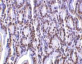
Immunohistochemistry (Formalin/PFA-fixed paraffin-embedded sections) - Anti-PHAP1 antibody (ab5991)
ab5991 at 2µg/ml staining PHAP1 in mouse small intestine tissue by IHC

Western blot - Anti-PHAP1 antibody (ab5991)
All lanes : Anti-PHAP1 antibody (ab5991) at 1 µg/ml
Lane 1 : Mouse heart tissue lysate
Lane 2 : Mouse pancreas tissue lysate
Lane 3 : Mouse testis tissue lysate
Lane 4 : Mouse liver tissue lysate
Lane 5 : Mouse spleen tissue lysate
Lane 6 : Mouse kidney tissue lysate
Lane 7 : Mouse colon tissue lysate
Lane 8 : Mouse stomach tissue lysate
Lane 9 : Mouse brain tissue lysate
Lane 10 : Mouse skin tissue lysate
Lane 11 : Mouse skeletal muscle tissue lysate
Lane 12 : Mouse bladder tissue lysate
Lane 13 : Mouse lung tissue lysate
Lane 14 : K562 cell lysate
Lysates/proteins at 15 µg/ml per lane.
Secondary
All lanes : HRP-conjugated goat anti-rabbit IgG at 1/10000 dilution
Incubated with the primary antibody for 1 hour at room temperature in 5% NFDM/TBST.
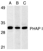
Western blot - Anti-PHAP1 antibody (ab5991)
Western blot analysis of PHAP I expression in human Raji cell (A), mouse (B) and rat (C) testis tissue llysates with anti-PHAP I at 1 µg/ml.

Immunocytochemistry/ Immunofluorescence - Anti-PHAP1 antibody (ab5991)
Immunofluorescence of PHAP I in mouse small intestine cells using ab5991 at 20µg/ml.


