Anti-eIF4E (phospho S209)抗体
参阅全部 eIF4E 一抗
兔多克隆抗体to eIF4E (phospho S209)
Rabbit
This phosphorylation site specific antibody is selective for eIF-4E containing a phosphate on serine 209.
适用于: ICC/IF, WBmore details
与反应: Rat, Human
预测可用于: Mouse, Rabbit, a wide range of other species![]()
Synthetic peptide corresponding to Human eIF4E (phospho S209).
WB: HeLa , HEK293, L6 cell lysate ICC/IF: U-87 MG cells
The Life Science industry has been in the grips of a reproducibility crisis for a number of years. Abcam is leading the way in addressing this with our range of recombinant monoclonal antibodies and knockout edited cell lines for gold-standard validation. Please check that this product meets your needs before purchasing.
If you have any questions, special requirements or concerns, please send us an inquiry and/or contact our Support team ahead of purchase. Recommended alternatives for this product can be found below, along with publications, customer reviews and Q&As
Liquid
Shipped at 4°C. Upon delivery aliquot and store at -20°C or -80°C. Avoid repeated freeze / thaw cycles.
pH: 7.30
Preservative: 0.05% Sodium azide
Constituents: PBS, 50% Glycerol (glycerin, glycerine), 1% BSA
Immunogen affinity purified
Purified from rabbit serum by sequential epitope-specific chromatography. The antibody has been negatively preadsorbed using a non-phosphopeptide corresponding to the site of phosphorylation to remove antibody that is reactive with non-phosphorylated eIF-4E. The final product is generated by affinity chromatography using an eIF-4E-derived peptide that is phosphorylated at serine 209.
多克隆
IgG
Abpromise™承诺保证使用ab4774于以下的经测试应用
“应用说明”部分 下显示的仅为推荐的起始稀释度;实际最佳的稀释度/浓度应由使用者检定。
| 应用 | Ab评论 | 说明 |
|---|---|---|
| ICC/IF | 1/250. | |
| WB | 1/1000. Detects a band of approximately 25 kDa (predicted molecular weight: 25 kDa). |
Entrez Gene: 1977 Human
Entrez Gene: 13684 Mouse
Omim: 133440 Human
SwissProt: P06730 Human
SwissProt: P63073 Mouse
SwissProt: P29338 Rabbit
Unigene: 249718 Human
Unigene: 3941 Mouse
Unigene: 11275 Rat
AUTS19 antibody
CBP antibody
eIF 4E antibody
eIF 4F 25 kDa subunit antibody
EIF 4F antibody
eIF-4E antibody
eIF-4F 25 kDa subunit antibody
eIF4E antibody
EIF4E1 antibody
EIF4EL1 antibody
EIF4F antibody
Eukaryotic translation initiation factor 4 E antibody
Eukaryotic translation initiation factor 4E antibody
Eukaryotic translation initiation factor 4E like 1 antibody
IF4E_HUMAN antibody
Messanger RNA Cap Binding Protein eIF 4E antibody
MGC111573 antibody
mRNA cap binding protein antibody
mRNA cap-binding protein antibody
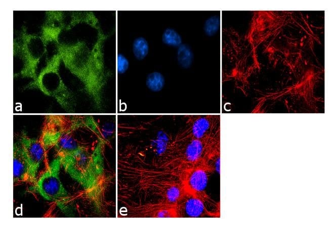
Immunocytochemistry/ Immunofluorescence - Anti-eIF4E (phospho S209) antibody (ab4774)
Immunofluorescence analysis using 70% confluent log phase U-87 MG cells labeled for Phospho-eIF4E pSer209 using ab4774. The cells were fixed with 4% paraformaldehyde for 10 minutes, permeabilized with 0.1% Triton™ X-100 for 10 minutes, and blocked with 1% BSA for 1 hour at room temperature. The cells were labeled with ab4774 at 1:250 dilution in 0.1% BSA and incubated for 3 hours at room temperature and then labeled with Goat anti-Rabbit IgG (H+L) Superclonal™ Secondary Antibody, Alexa Fluor® 488 conjugate at a dilution of 1:2000 for 45 minutes at room temperature (Panel A: green). Nuclei (Panel B: blue) were stained with SlowFade® Gold Antifade Mountant with DAPI. F-actin (Panel C: red) was stained with Rhodamine Phalloidin (1:300). Panel D is a merged image showing cytoplasmic localization. Panel E is a no primary antibody control. The images were captured at 60X magnification.

Western blot - Anti-eIF4E (phospho S209) antibody (ab4774)
All lanes : Anti-eIF4E (phospho S209) antibody (ab4774)
Lane 1 : HeLa whole cell extracts
Lane 2 : Serum starved HeLa
Lane 3 : HeLa Serum Starved for overnight followed by Serum Released
Lane 4 : HEK-293
Lane 5 : HEK-293 treated for 30 minutes with 25 µg/mL of Anisomycin
Lane 6 : L6
Lane 7 : L6 treated for 10 minutes with 200 ng/mL of Insulin
Lysates/proteins at 20 µg per lane.
Secondary
All lanes : Goat anti-Rabbit IgG (H+L) Superclonal™ Secondary Antibody, HRP conjugate at 0.4 µg/ml
Predicted band size: 25 kDa
Western blot analysis was performed on HeLa, HEK-293 (human cell lines) and L6 (rat cell line) cell lysates with various treatments, blotted for eIF4E (pS209) using ab4774. Two bands ~ 25 and 28 kDa band corresponding to eIF4E (pS209) was observed across cell lines tested. Known quantity of protein samples were electrophoresed using Novex® NuPAGE® 12 % Bis-Tris gel, XCell SureLock™ Electrophoresis System and Novex® Sharp Pre-Stained Protein Standard. Resolved proteins were then transferred onto a nitrocellulose membrane with iBlot® 2 Dry Blotting System. The membrane was probed with the relevant primary and secondary Antibody following blocking with 5 % skimmed milk. Chemiluminescent detection was performed using Pierce™ ECL Western Blotting Substrate

Western blot - Anti-eIF4E (phospho S209) antibody (ab4774)
Extracts of HeLa cells were resolved by SDS-PAGE on a 4-20% Tris-glycine gel and transferred to PVDF. The membrane was blocked with a 5% BSA-TBST buffer for one hour at room temperature, either left untreated (1-4) or treated with lambda (?) phosphatase (5), and then incubated with the eIF4E (phospho S209) antibody for two hours at room temperature in a 3% BSA-TBST buffer, following prior incubation with: no peptide (1,5), the nonphosphopeptide corresponding to the phosphopeptide immunogen (2), a generic phosphoserine-containing peptide (3), or the phosphopeptide immunogen (4). After washing, the membrane was incubated with goat F(ab’)2 anti-rabbit IgG alkaline phosphatase and signals were detected using the Pierce SuperSignal™ method.
The data show that only the peptide corresponding to eIF4E (phospho S209) blocks the antibody signal, demonstrating the specificity of the antibody. The data also show that phosphatase stripping eliminates the signal, verifying that the antibody i


