| 存储条件 |
|---|
| Product Name | Noggin Recombinant Rabbit Monoclonal Antibody |
|---|---|
| Antibody Type | Primary Antibodies |
| Immunogen | Recombinant protein within human Noggin aa 28-232. |
| Clonality | monoclonal |
|---|---|
| Isotype | IgG |
| Host Species | Rabbit |
| Tested Applications | IHCWB |
| WB:1:2000 IHC:1:50000 | |
| Species Reactivity | HumanMouseRat |
| Concentration | 1mg/ml |
| Purification | Protein A |
| Gene Symbol | NOG |
|---|---|
| Gene Synonyms | SYM1 SYNS1 SYNS1A |
| Gene Full Name | noggin |
| Gene Summary | The secreted polypeptide, encoded by this gene, binds and inactivates members of the transforming growth factor-beta (TGF-beta) superfamily signaling proteins, such as bone morphogenetic protein-4 (BMP4). By diffusing through extracellular matrices more efficiently than members of the TGF-beta superfamily, this protein may have a principal role in creating morphogenic gradients. The protein appears to have pleiotropic effect, both early in development as well as in later stages. It was originally isolated from Xenopus based on its ability to restore normal dorsal-ventral body axis in embryos that had been artificially ventralized by UV treatment. The results of the mouse knockout of the ortholog suggest that it is involved in numerous developmental processes, such as neural tube fusion and joint formation. Recently, several dominant human NOG mutations in unrelated families with proximal symphalangism (SYM1) and multiple synostoses syndrome (SYNS1) were identified; both SYM1 and SYNS1 have multiple joint fusion as their principal feature, and map to the same region (17q22) as this gene. All of these mutations altered evolutionarily conserved amino acid residues. The amino acid sequence of this human gene is highly homologous to that of Xenopus, rat and mouse. [provided by RefSeq, Jul 2008] |
| Alternative Names | Nog antibody NOGG_HUMAN antibody Noggin antibody SYM 1 antibody SYM1 antibody Symphalangism 1 (proximal) antibody Synostoses (multiple) syndrome 1 antibody SYNS 1 antibody SYNS1 antibody |
| Molecular Weight(MW) | 26kDa(Observed band size: 23kDa) |
| Cellular Localization | Secreted. |
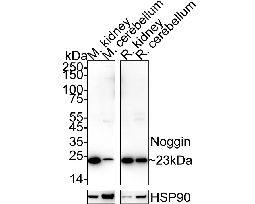
WB
Western blot analysis of Noggin on different lysates with Rabbit anti-Noggin antibody at 1/2,000 dilution. Lane 1: Mouse kidney tissue lysate, Lane 2: Mouse cerebellum tissue lysate, Lane 3: Rat kidney tissue lysate, Lane 4: Rat cerebellum tissue lysate, Lysates/proteins at 20 µg/Lane. Exposure time: 1 minute; 4-20% SDS-PAGE gel. Proteins were transferred to a PVDF membrane and blocked with 5% NFDM/TBST for 1 hour at room temperature. The primary antibody at 1/2,000 dilution was used in 5% NFDM/TBST at 4℃ overnight. Goat Anti-Rabbit IgG - HRP Secondary Antibody at 1/50,000 dilution was used for 1 hour at room temperature.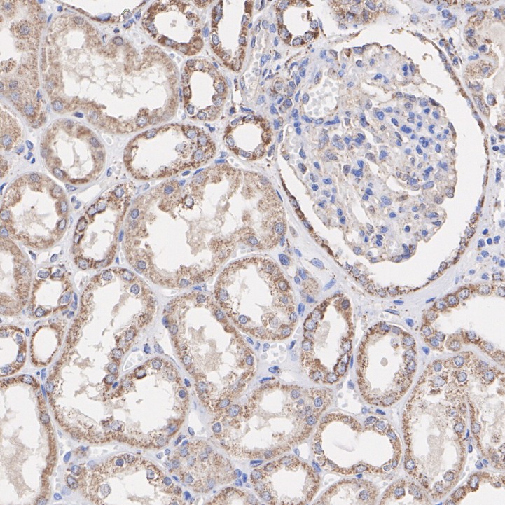
IHC
Immunohistochemical analysis of paraffin-embedded human kidney tissue with Rabbit anti-Noggin antibody at 1/50,000 dilution. The section was pre-treated using heat mediated antigen retrieval with Tris-EDTA buffer (pH 9.0) for 20 minutes. The tissues were blocked in 1% BSA for 20 minutes at room temperature, washed with ddH2O and PBS, and then probed with the primary antibody at 1/50,000 dilution for 1 hour at room temperature. The detection was performed using an HRP conjugated compact polymer system. DAB was used as the chromogen. Tissues were counterstained with hematoxylin and mounted with DPX.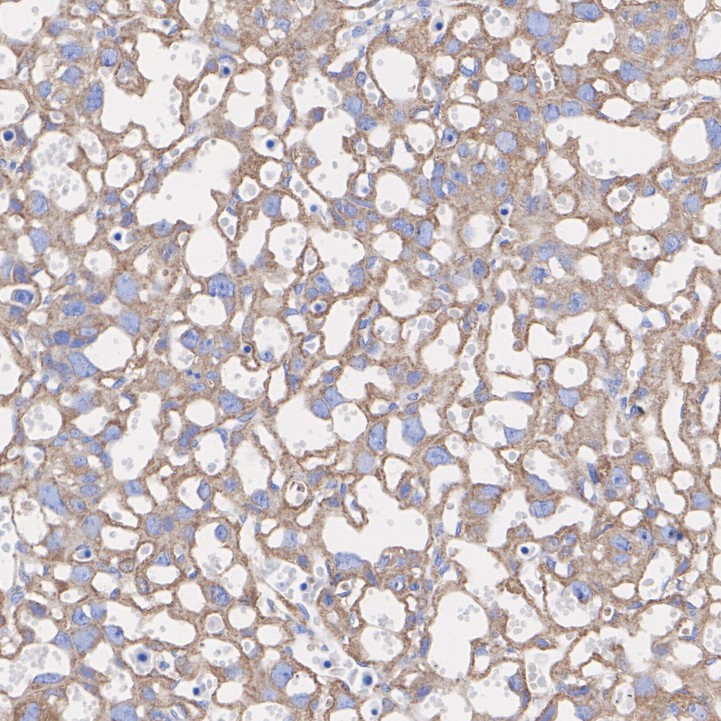
IHC
Immunohistochemical analysis of paraffin-embedded mouse placenta tissue with Rabbit anti-Noggin antibody at 1/50,000 dilution. The section was pre-treated using heat mediated antigen retrieval with Tris-EDTA buffer (pH 9.0) for 20 minutes. The tissues were blocked in 1% BSA for 20 minutes at room temperature, washed with ddH2O and PBS, and then probed with the primary antibody at 1/50,000 dilution for 1 hour at room temperature. The detection was performed using an HRP conjugated compact polymer system. DAB was used as the chromogen. Tissues were counterstained with hematoxylin and mounted with DPX.| Application Notes | WB:1:2000 IHC:1:50000 |
|---|
| Form | Liquid |
|---|---|
| Storage Instructions | Store at +4℃ after thawing. Aliquot store at -20℃. Avoid repeated freeze / thaw cycles. |
| Storage Buffer | 1*TBS (pH7.4), 0.05% BSA, 40% Glycerol. Preservative: 0.05% Sodium Azide. |
抱歉,暂无相关文献







