| 存储条件 |
|---|
| Product Name | E-Cadherin Recombinant Rabbit Monoclonal Antibody |
|---|---|
| Antibody Type | Primary Antibodies |
| Immunogen | Synthetic peptide within human E-Cadherin aa 580-630 (Extracellular). |
| Clonality | monoclonal |
|---|---|
| Isotype | IgG |
| Host Species | Rabbit |
| Tested Applications | FCICC/IFIF-PIHCWB |
| WB:1:5000 IHC:1:200 ICC/IF:1:2000 IF-P:1:50-1:200 FC:1:1000 | |
| Species Reactivity | Human |
| Concentration | 1mg/ml |
| Purification | Protein A |
| Gene Symbol | CDH1 |
|---|---|
| Gene Synonyms | UVO CDHE ECAD LCAM Arc-1 BCDS1 CD324 |
| Gene Full Name | cadherin 1 |
| Gene Summary | A large family of ATPases has been described, whose key feature is that they share a conserved region of about 220 amino acids that contains an ATP-binding site. The proteins that belong to this family either contain one or two AAA (ATPases Associated with diverse cellular Activities) domains. AAA family proteins often perform chaperone-like functions that assist in the assembly, operation, or disassembly of protein complexes. The protein encoded by this gene contains two AAA domains, as well as a bromodomain. [provided by RefSeq, Jul 2008] |
| Alternative Names | Arc 1 antibody CADH1_HUMAN antibody Cadherin 1 antibody cadherin 1 type 1 E-cadherin antibody Cadherin1 antibody CAM 120/80 antibody CD 324 antibody CD324 antibody CD324 antigen antibody cdh1 antibody Arc 1 antibody CADH1_HUMAN antibody Cadherin 1 antibody cadherin 1 type 1 E-cadherin antibody Cadherin1 antibody CAM 120/80 antibody CD 324 antibody CD324 antibody CD324 antigen antibody cdh1 antibody CDHE antibody E-Cad/CTF3 antibody E Cadherin antibody E-cadherin antibody ECAD antibody Epithelial cadherin antibody epithelial calcium dependant adhesion protein antibody LCAM antibody Liver cell adhesion molecule antibody UVO antibody Uvomorulin antibody |
| Molecular Weight(MW) | 97kDa(Observed band size:80-120kDa) |
| Cellular Localization | Cell junction, Cell membrane, Endosome, Golgi apparatus. |
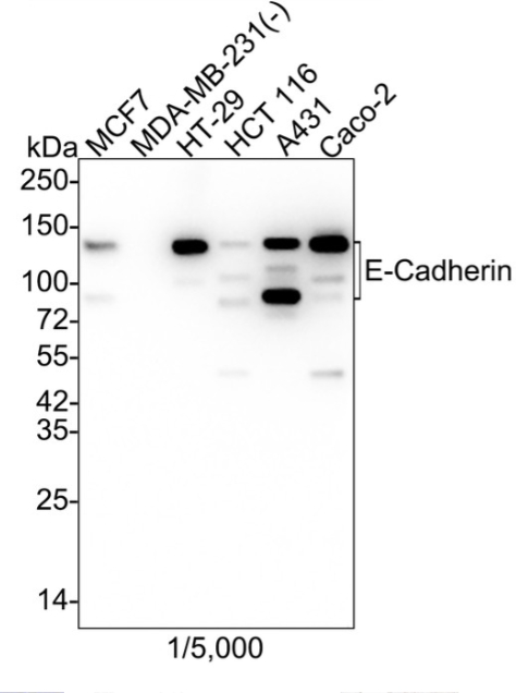
WB
Western blot analysis of E-Cadherin on different lysates with Rabbit anti-E-Cadherin antibody at 1/5,000 dilution. Lane 1: MCF7 cell lysate, Lane 2: MDA-MB-231 cell lysate (negative), Lane 3: HT-29 cell lysate, Lane 4: HCT 116 cell lysate, Lane 5: A431 cell lysate, Lane 6: Caco-2 cell lysate, Lysates/proteins at 20 µg/Lane. Exposure time: 43 seconds; 4-20% SDS-PAGE gel. Proteins were transferred to a PVDF membrane and blocked with 5% NFDM/TBST for 1 hour at room temperature. The primary antibody at 1/5,000 dilution was used in 5% NFDM/TBST at 4℃ overnight. Goat Anti-Rabbit IgG - HRP Secondary Antibody at 1/50,000 dilution was used for 1 hour at room temperature.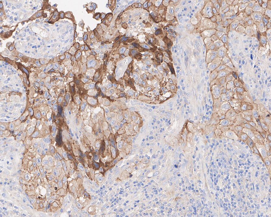
IHC
Immunohistochemical analysis of paraffin-embedded human lung carcinoma tissue with Rabbit anti-E-Cadherin antibody at 1/200 dilution. The section was pre-treated using heat mediated antigen retrieval with Tris-EDTA buffer (pH 9.0) for 20 minutes. The tissues were blocked in 1% BSA for 20 minutes at room temperature, washed with ddH2O and PBS, and then probed with the primary antibody at 1/200 dilution for 1 hour at room temperature. The detection was performed using an HRP conjugated compact polymer system. DAB was used as the chromogen. Tissues were counterstained with hematoxylin and mounted with DPX.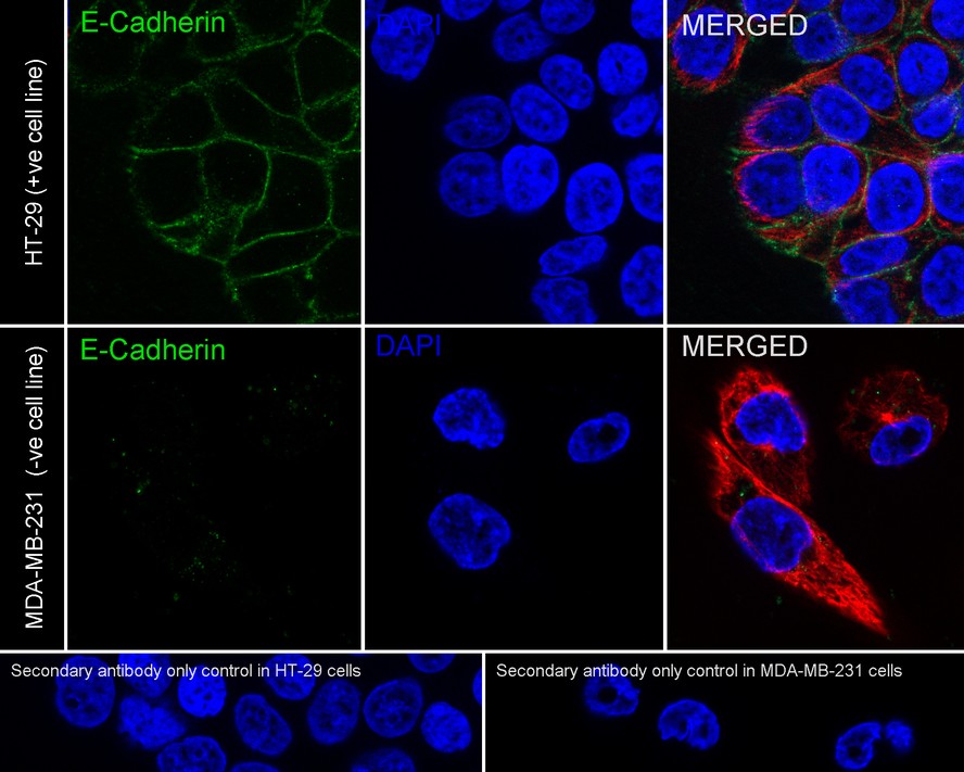
ICC/IF
Immunocytochemistry analysis of HT-29 (positive) and MDA-MB-231 (negative) cells labeling E-Cadherin with Rabbit anti-E-Cadherin antibody at 1/2,000 dilution. Cells were fixed in 4% paraformaldehyde for 20 minutes at room temperature, permeabilized with 0.1% Triton X-100 in PBS for 5 minutes at room temperature, then blocked with 1% BSA in 10% negative goat serum for 1 hour at room temperature. Cells were then incubated with Rabbit anti-E-Cadherin antibody at 1/2,000 dilution in 1% BSA in PBST overnight at 4 ℃. Goat Anti-Rabbit IgG H&L (488) was used as the secondary antibody at 1/1,000 dilution. PBS instead of the primary antibody was used as the secondary antibody only control. Nuclear DNA was labelled in blue with DAPI. Beta tubulin (red) was stained at 1/100 dilution overnight at +4℃. Goat Anti-Mouse IgG H&L (594) was used as the secondary antibody at 1/1,000 dilution.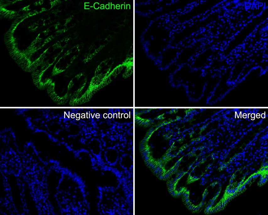
IF-P
Immunofluorescence analysis of paraffin-embedded human colon tissue labeling E-Cadherin with Rabbit anti-E-Cadherin antibody at 1/50 dilution. The section was pre-treated using heat mediated antigen retrieval with Tris-EDTA buffer (pH 9.0) for 20 minutes. The tissues were blocked in 10% negative goat serum for 1 hour at room temperature, washed with PBS, and then probed with the primary antibody (reen) at 1/50 dilution overnight at 4 ℃, washed with PBS. Goat Anti-Rabbit IgG H&L (488) was used as the secondary antibody at 1/1,000 dilution. Nuclei were counterstained with Hoechst 33258 (blue).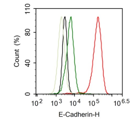
FC
Flow cytometric analysis of HT-29 (positive, red) and MDA-MB-231 (negative, green) cells labeling E-Cadherin. Cells were fixed and permeabilized. Then stained with the primary antibody (red) at 1/1,000 dilution, compared with Rabbit IgG Isotype Control (HT-29 black, MDA-MB-231 light green). After incubation of the primary antibody at +4℃ for an hour, the cells were stained with a 488 conjugate-Goat anti-Rabbit IgG Secondary antibody at 1/1,000 dilution for 30 minutes at +4℃.| Application Notes | WB:1:5000 IHC:1:200 ICC/IF:1:2000 IF-P:1:50-1:200 FC:1:1000 |
|---|
| Form | Liquid |
|---|---|
| Storage Instructions | Store at +4℃ after thawing. Aliquot store at -20℃. Avoid repeated freeze / thaw cycles. |
| Storage Buffer | 1*TBS (pH7.4), 0.05% BSA, 40% Glycerol. Preservative: 0.05% Sodium Azide. |
抱歉,暂无相关文献







