| 存储条件 |
|---|
| Product Name | CD11c Recombinant Rabbit Monoclonal Antibody |
|---|---|
| Antibody Type | Primary Antibodies |
| Immunogen | Synthetic peptide within Human CD11c aa 1,114-1,163 / 1,163. |
| Clonality | monoclonal |
|---|---|
| Isotype | IgG |
| Host Species | Rabbit |
| Tested Applications | IF-PIHC |
| IHC:1:400-1:1000 IF-P:1:1000 | |
| Species Reactivity | Human |
| Concentration | 1mg/ml |
| Purification | Protein A |
| Gene Symbol | Itgax |
|---|---|
| Gene Synonyms | CD11C SLEB6 |
| Gene Full Name | integrin subunit alpha X |
| Gene Summary | This gene encodes the integrin alpha X chain protein. Integrins are heterodimeric integral membrane proteins composed of an alpha chain and a beta chain. This protein combines with the beta 2 chain (ITGB2) to form a leukocyte-specific integrin referred to as inactivated-C3b (iC3b) receptor 4 (CR4). The alpha X beta 2 complex seems to overlap the properties of the alpha M beta 2 integrin in the adherence of neutrophils and monocytes to stimulated endothelium cells, and in the phagocytosis of complement coated particles. Two transcript variants encoding different isoforms have been found for this gene. [provided by RefSeq, Nov 2013] |
| Alternative Names | 95 alpha chain antibody 95 antibody CD 11c antibody CD11 antigen-like family member C antibody CD11c antibody CD11c antigen antibody Complement component 3 receptor 4 subunit antibody CR4 antibody Integrin alpha X antibody Integrin alpha X chain antibody 95 alpha chain antibody 95 antibody CD 11c antibody CD11 antigen-like family member C antibody CD11c antibody CD11c antigen antibody Complement component 3 receptor 4 subunit antibody CR4 antibody Integrin alpha X antibody Integrin alpha X chain antibody Integrin alpha-X antibody Integrin aX antibody Integrin subunit alpha X antibody integrin alpha X (antigen CD11C (p150) alpha polypeptide) antibody integrin alpha X (complement component 3 receptor 4 subunit antibody ITAX_HUMAN antibody ITGAX antibody LEU M5 alpha subunit antibody Leu M5 antibody Leukocyte adhesion glycoprotein p150 95 alpha chain antibody Leukocyte adhesion glycoprotein p150 antibody Leukocyte adhesion receptor p150 95 antibody Leukocyte adhesion receptor p150 antibody Leukocyte surface antigen p150 95 alpha subunit antibody Leukocyte surface antigen p150 alpha subunit antibody Myeloid membrane antigen alpha subunit antibody p150 95 integrin alpha chain antibody p150 antibody p150/95 antibody SLEB6 antibody |
| Molecular Weight(MW) | 129kDa |
| Cellular Localization | Membrane |
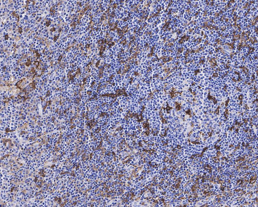
IHC
Immunohistochemical analysis of paraffin-embedded human Hodgkin's lymphoma tissue with Rabbit anti-CD11c antibody at 1/800 dilution. The section was pre-treated using heat mediated antigen retrieval with Tris-EDTA buffer (pH 9.0) for 20 minutes. The tissues were blocked in 1% BSA for 20 minutes at room temperature, washed with ddH2O and PBS, and then probed with the primary antibody at 1/800 dilution for 1 hour at room temperature. The detection was performed using an HRP conjugated compact polymer system. DAB was used as the chromogen. Tissues were counterstained with hematoxylin and mounted with DPX.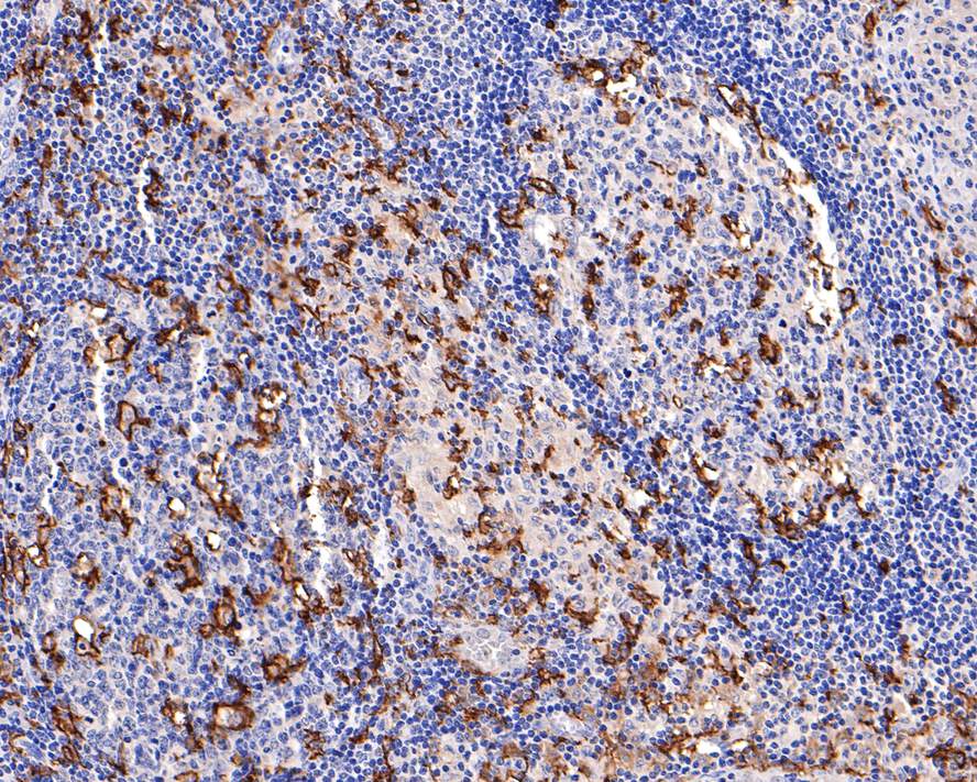
IHC
Immunohistochemical analysis of paraffin-embedded human lymph nodes tissue with Rabbit anti-CD11c antibody at 1/400 dilution. The section was pre-treated using heat mediated antigen retrieval with Tris-EDTA buffer (pH 9.0) for 20 minutes. The tissues were blocked in 1% BSA for 20 minutes at room temperature, washed with ddH2O and PBS, and then probed with the primary antibody at 1/400 dilution for 1 hour at room temperature. The detection was performed using an HRP conjugated compact polymer system. DAB was used as the chromogen. Tissues were counterstained with hematoxylin and mounted with DPX.
IF-P
Fluorescence multiplex immunohistochemical analysis of human tonsil (Formalin/PFA-fixed paraffin-embedded sections). The merged image of anti-CD45 (Yellow), anti-CD15 (Green) and anti-CD11c (Red) on tonsil. HRP Conjugated UltraPolymer Goat Polyclonal Antibody HA1119/HA1120 was used as a secondary antibody. The immunostaining was performed with the Sequential Immuno-staining Kit. The section was incubated in three rounds of staining: in the order of anti-CD45 (1/500 dilution), anti-CD15 (1/500 dilution) and anti-CD11c (1/1,000 dilution) for 20 mins at room temperature. Each round was followed by a separate fluorescent tyramide signal amplification system. Heat mediated antigen retrieval with Tris-EDTA buffer (pH 9.0) for 30 mins at 95℃. DAPI (blue) was used as a nuclear counter stain. Image acquisition was performed with Zeiss Observer 7 Inverted Fluorescence Microscope.| Application Notes | IHC:1:400-1:1000 IF-P:1:1000 |
|---|
| Form | Liquid |
|---|---|
| Storage Instructions | Store at +4℃ after thawing. Aliquot store at -20℃. Avoid repeated freeze / thaw cycles. |
| Storage Buffer | PBS (pH7.4), 0.1% BSA, 40% Glycerol. Preservative: 0.05% Sodium Azide. |
抱歉,暂无相关文献







