| 存储条件 |
|---|
| Product Name | c-Fos Recombinant Rabbit Monoclonal Antibody |
|---|---|
| Antibody Type | Primary Antibodies |
| Product description | Protein c-Fos is a proto-oncogene that is the human homolog of the retroviral oncogene vfos. It is encoded in humans by the FOS gene. It was first discovered in rat fibroblasts as the transforming gene of the FBJ MSV (Finkel–Biskis–Jinkins murine osteogenic sarcoma virus). It is a part of a bigger Fos family of transcription factors which includes c-Fos, FosB, Fra-1 and Fra-2. It has been mapped to chromosome region 14q21→q31. c-Fos encodes a 62 kDa protein, which forms heterodimer with c-jun (part of Jun family of transcription factors), resulting in the formation of AP-1 (Activator Protein-1) complex which binds DNA at AP-1 specific sites at the promoter and enhancer regions of target genes and converts extracellular signals into changes of gene expression. It plays an important role in many cellular functions and has been found to be overexpressed in a variety of cancers. |
| Immunogen | Recombinant protein within human Protein c-Fos aa 1-380 |
| Clonality | monoclonal |
|---|---|
| Isotype | IgG |
| Host Species | Rabbit |
| Tested Applications | ICC/IFIHCWB |
| WB:1:500-1:2000 IHC:1:200-1:1000 ICC:1:50-1:200 | |
| Species Reactivity | HumanMouseRat |
| Concentration | 1mg/ml |
| Purification | Protein A |
| Gene Symbol | FOS |
|---|---|
| Gene Synonyms | AP-1 C-FOS p55 |
| Gene Full Name | Fos proto-oncogene, AP-1 transcription factor subunit |
| Gene Summary | The Fos gene family consists of 4 members: FOS, FOSB, FOSL1, and FOSL2. These genes encode leucine zipper proteins that can dimerize with proteins of the JUN family, thereby forming the transcription factor complex AP-1. As such, the FOS proteins have been implicated as regulators of cell proliferation, differentiation, and transformation. In some cases, expression of the FOS gene has also been associated with apoptotic cell death. [provided by RefSeq, Jul 2008] |
| Alternative Names | Activator protein 1 antibody AP 1 antibody C FOS antibody Cellular oncogene c fos antibody Cellular oncogene fos antibody FBJ murine osteosarcoma viral (v fos) oncogene homolog (oncogene FOS) antibody FBJ murine osteosarcoma viral oncogene homolog antibody FBJ murine osteosarcoma viral v fos oncogene homolog antibody FBJ Osteosarcoma Virus antibody FOS antibody Activator protein 1 antibody AP 1 antibody C FOS antibody Cellular oncogene c fos antibody Cellular oncogene fos antibody FBJ murine osteosarcoma viral (v fos) oncogene homolog (oncogene FOS) antibody FBJ murine osteosarcoma viral oncogene homolog antibody FBJ murine osteosarcoma viral v fos oncogene homolog antibody FBJ Osteosarcoma Virus antibody FOS antibody FOS protein antibody FOS_HUMAN antibody G0 G1 switch regulatory protein 7 antibody G0/G1 switch regulatory protein 7 antibody G0S7 antibody Oncogene FOS antibody p55 antibody proto oncogene c Fos antibody Proto oncogene protein c fos antibody Proto-oncogene c-Fos antibody v fos FBJ murine osteosarcoma viral oncogene homolog antibody |
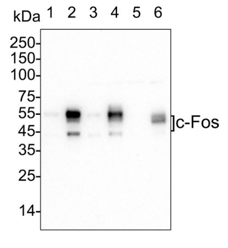
| Molecular Weight(MW) | 41-55 kDa |
| Cellular Localization | Nucleus, Endoplasmic reticulum, Cytoplasm, cytosol. |
WB
Western blot analysis of c-Fos on different lysates with Rabbit anti-c-Fos antibody at 1/1,000 dilution . Lane 1: HeLa cell lysate, Lane 2: HeLa serum starved for 40 hours then add 20% FBS for 2 hours cell lysate, Lane 3: RAW264.7 cell lysate, Lane 4: RAW264.7 serum starved for 16 hours then add 200nM PMA for 4 hours cell lysate, Lane 5: C6 cell lysate, Lane 6: C6 serum starved for 16 hours then add 10% FBS for 30 minutes cell lysate, Lysates/proteins at 20 µg/Lane.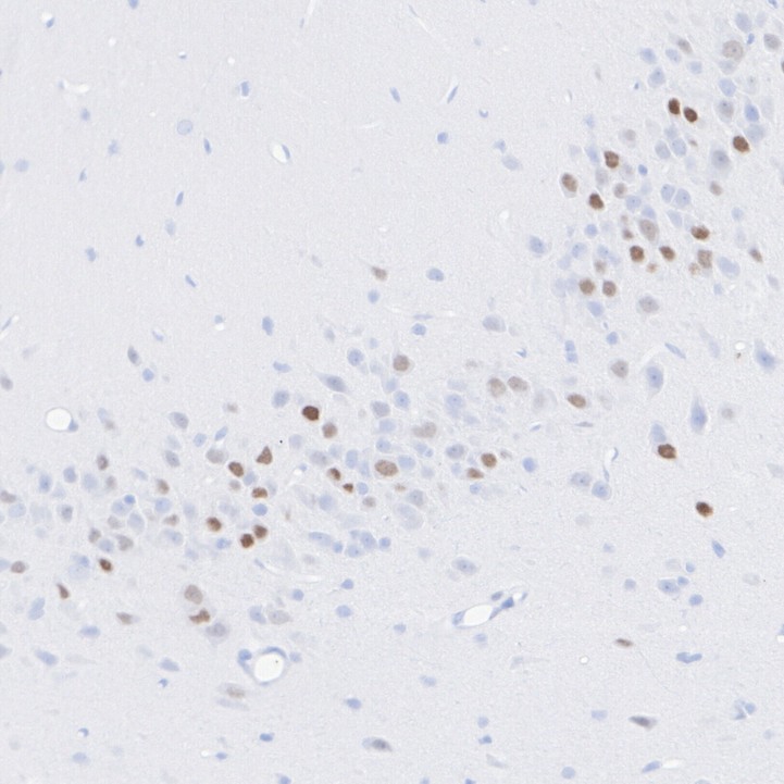
IHC
Immunohistochemical analysis of paraffin-embedded rat brain (piriform area) tissue with Rabbit anti-c-Fos antibody at 1/500 dilution. The section was pre-treated using heat mediated antigen retrieval with sodium citrate buffer (pH 6.0) for 2 minutes. The tissues were blocked in 1% BSA for 20 minutes at room temperature, washed with ddH2O and PBS, and then probed with the primary antibody at 1/500 dilution for 1 hour at room temperature. The detection was performed using an HRP conjugated compact polymer system. DAB was used as the chromogen. Tissues were counterstained with hematoxylin and mounted with DPX.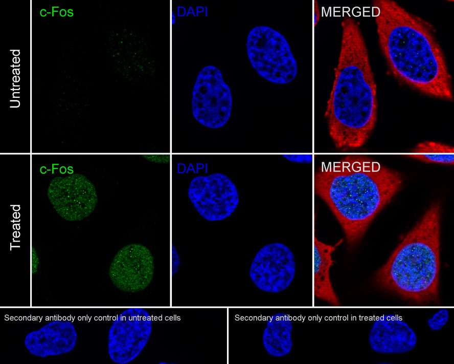
ICC/IF
Immunocytochemistry analysis of HeLa cells serum starved for 40 hours then add 20% FBS for 2 hours labeling c-Fos with Rabbit anti-c-Fos antibody at 1/100 dilution. Cells were fixed in 4% paraformaldehyde for 20 minutes at room temperature, permeabilized with 0.1% Triton X-100 in PBS for 5 minutes at room temperature, then blocked with 1% BSA in 10% negative goat serum for 1 hour at room temperature. Cells were then incubated with Rabbit anti-c-Fos antibody at 1/100 dilution in 1% BSA in PBST overnight at 4 ℃. Goat Anti-Rabbit IgG H&L (iFluor™ 488) was used as the secondary antibody at 1/1,000 dilution. PBS instead of the primary antibody was used as the secondary antibody only control. Nuclear DNA was labelled in blue with DAPI. Beta tubulin ( red) was stained at 1/100 dilution overnight at +4℃. Goat Anti-Mouse IgG H&L (iFluor™ 594) was used as the secondary antibody at 1/1,000 dilution.| Application Notes | WB:1:500-1:2000 IHC:1:200-1:1000 ICC:1:50-1:200 |
|---|
| Form | Liquid |
|---|---|
| Storage Instructions | Shipped at 4°C. Store at +4°C short term (1-2 weeks). Store at -20°C long term. Avoid freeze / thaw cycle. |
| Storage Buffer | PBS (pH7.4), 0.1% BSA, 40% Glycerol. Preservative: 0.05% Sodium Azide |
抱歉,暂无相关文献







