| 存储条件 |
|---|
| Product Name | Phospho-POLR2A (S2) Recombinant Rabbit Monoclonal Antibody |
|---|---|
| Antibody Type | Primary Antibodies |
| Immunogen | Synthetic phospho-peptide corresponding to residues surrounding Ser2 of Human POLR2A aa 1590-1630 / 1970. |
| Modification | p-S2 |
| Clonality | monoclonal |
|---|---|
| Isotype | IgG |
| Host Species | Rabbit |
| Tested Applications | FCICC/IFIHCWB |
| WB:1:500-1:2000 IHC:1:50-1:200 ICC:1:100-1:500 FC:1:50-1:100 | |
| Species Reactivity | HumanMouseRat |
| Concentration | 1mg/ml |
| Purification | Protein A |
| Gene Symbol | POLR2A |
|---|---|
| Gene Synonyms | RPB1 RPO2 POLR2 POLRA RPBh1 RPOL2 NEDHIB RpIILS hsRPB1 hRPB220 |
| Gene Full Name | RNA polymerase II subunit A |
| Gene Summary | This gene encodes the largest subunit of RNA polymerase II, the polymerase responsible for synthesizing messenger RNA in eukaryotes. The product of this gene contains a carboxy terminal domain composed of heptapeptide repeats that are essential for polymerase activity. These repeats contain serine and threonine residues that are phosphorylated in actively transcribing RNA polymerase. In addition, this subunit, in combination with several other polymerase subunits, forms the DNA binding domain of the polymerase, a groove in which the DNA template is transcribed into RNA. [provided by RefSeq, Jul 2008] |
| Target | The tandem heptapeptide repeats in the C-terminal domain (CTD) can be highly phosphorylated. The phosphorylation activates Pol II. Phosphorylation occurs mainly at residues 'Ser-2' and 'Ser-5' of the heptapeptide repeat and is mediated, at least, by CDK7 and CDK9. CDK7 phosphorylation of POLR2A associated with DNA promotes transcription initiation by triggering dissociation from DNA. Phosphorylation also takes place at 'Ser-7' of the heptapeptide repeat, which is required for efficient transcription of snRNA genes and processing of the transcripts. The phosphorylation state is believed to result from the balanced action of site-specific CTD kinases and phosphatases, and a 'CTD code' that specifies the position of Pol II within the transcription cycle has been proposed. Dephosphorylated by the protein phosphatase CTDSP1.; Among tandem heptapeptide repeats of the C-terminal domain (CTD) some do not match the Y-S-P-T-S-P-S consensus, the seventh serine residue 'Ser-7' being replaced by a lysine. 'Lys-7' in these non-consensus heptapeptide repeats can be alternatively acetylated, methylated and dimethylated. EP300 is one of the enzyme able to acetylate 'Lys-7'. Acetylation at 'Lys-7' of non-consensus heptapeptide repeats is associated with 'Ser-2' phosphorylation and active transcription. Regulates initiation or early elongation steps of transcription specially for inducible genes.; Methylated at Arg-1810 prior to transcription initiation when the CTD is hypophosphorylated, phosphorylation at Ser-1805 and Ser-1808 preventing this methylation. Symmetrically or asymmetrically dimethylated at Arg-1810 by PRMT5 and CARM1 respectively. Symmetric or asymmetric dimethylation modulates interactions with CTD-binding proteins like SMN1/SMN2 and TDRD3. SMN1/SMN2 interacts preferentially with the symmetrically dimethylated form while TDRD3 interacts with the asymmetric form. Through the recruitment of SMN1/SMN2, symmetric dimethylation is required for resolving RNA-DNA hybrids created by RNA polymerase II, that form R-loop in transcription terminal regions, an important step in proper transcription termination. CTD dimethylation may also facilitate the expression of select RNAs. Among tandem heptapeptide repeats of the C-terminal domain (CTD) some do not match the Y-S-P-T-S-P-S consensus, the seventh serine residue 'Ser-7' being replaced by a lysine. 'Lys-7' in these non-consensus heptapeptide repeats can be alternatively acetylated, methylated, dimethylated and trimethylated. Methylation occurs in the earliest transcription stages and precedes or is concomitant to 'Ser-5' and 'Ser-7' phosphorylation. Dimethylation and trimehtylation at 'Lys-7' of non-consensus heptapeptide repeats are exclusively associated with phosphorylated CTD.; Ubiquitinated by WWP2 leading to proteasomal degradation (By similarity). Following UV treatment, the elongating form of RNA polymerase II (RNA pol IIo) is ubiquitinated on UV damage sites without leading to degradation: ubiquitination is facilitated by KIAA1530/UVSSA and promotes RNA pol IIo backtracking to allow access to the nucleotide excision repair machinery. |
| Alternative Names | DNA directed RNA polymerase II A antibody DNA-directed RNA polymerase II largest subunit RNA polymerase II 220 kd subunit antibody DNA-directed RNA polymerase II subunit A antibody DNA-directed RNA polymerase II subunit RPB1 antibody DNA-directed RNA polymerase III largest subunit antibody hRPB220 antibody hsRPB1 antibody POLR2 antibody Polr2a antibody POLRA antibody DNA directed RNA polymerase II A antibody DNA-directed RNA polymerase II largest subunit RNA polymerase II 220 kd subunit antibody DNA-directed RNA polymerase II subunit A antibody DNA-directed RNA polymerase II subunit RPB1 antibody DNA-directed RNA polymerase III largest subunit antibody hRPB220 antibody hsRPB1 antibody POLR2 antibody Polr2a antibody POLRA antibody Polymerase (RNA) II (DNA directed) polypeptide A 220kDa antibody Polymerase (RNA) II (DNA directed) polypeptide A antibody RNA polymerase II subunit B1 antibody RNA-directed RNA polymerase II subunit RPB1 antibody RPB1 antibody RPB1_HUMAN antibody RPBh1 antibody RpIILS antibody RPO2 antibody RPOL2 antibody |
| Molecular Weight(MW) | 217kDa(Observed band size:260kDa) |
| Cellular Localization | Nucleus. |
WB
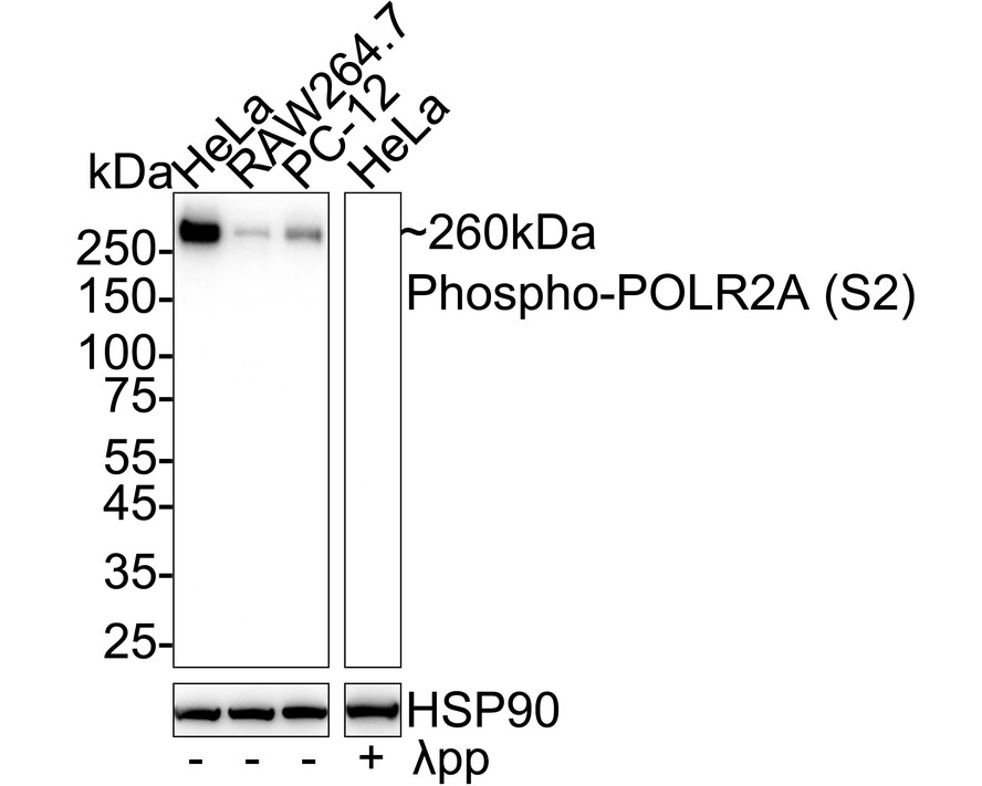
Western blot analysis of Phospho-POLR2A (S2) on different lysates with Rabbit anti-Phospho-POLR2A (S2) antibody at 1/1,000 dilution. Lane 1: HeLa cell lysate Lane 2: RAW264.7 cell lysate Lane 3: PC-12 cell lysate Lane 4: HeLa cell lysate, the membrane treated with λpp for 1 hour Lysates/proteins at 20 µg/Lane. Predicted band size: 217 kDa Observed band size: 260 kDa Exposure time: 6 seconds; 4-20% SDS-PAGE gel. Proteins were transferred to a PVDF membrane and blocked with 5% NFDM/TBST for 1 hour at room temperature. The primary antibody at 1/1,000 dilution was used in 5% NFDM/TBST at 4℃ overnight. Goat Anti-Rabbit IgG - HRP Secondary Antibody at 1/50,000 dilution was used for 1 hour at room temperature.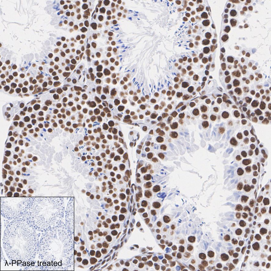
IHC
Immunohistochemical analysis of paraffin-embedded mouse testis tissue untreated / treated with λpp with Rabbit anti-Phospho-POLR2A (S2) antibody at 1/1,000 dilution. The section was pre-treated using heat mediated antigen retrieval with Tris-EDTA buffer (pH 9.0) for 20 minutes. The tissues were blocked in 1% BSA for 20 minutes at room temperature, washed with ddH2O and PBS, and then probed with the primary antibody at 1/1,000 dilution for 1 hour at room temperature. The detection was performed using an HRP conjugated compact polymer system. DAB was used as the chromogen. Tissues were counterstained with hematoxylin and mounted with DPX.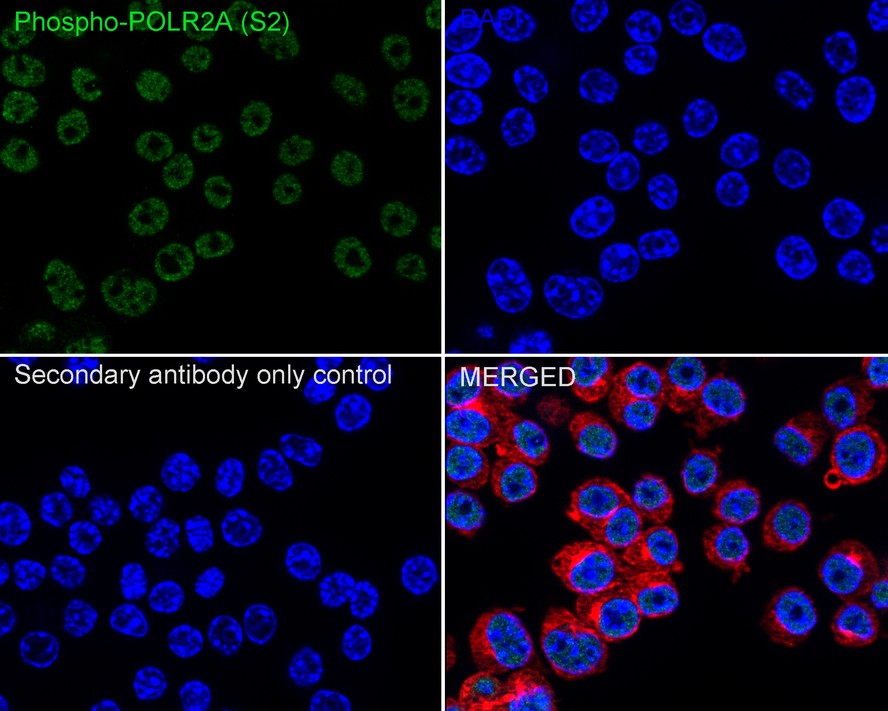
ICC/IF
Immunocytochemistry analysis of RAW264.7 cells labeling Phospho-POLR2A (S2) with Rabbit anti-Phospho-POLR2A (S2) antibody at 1/50 dilution. Cells were fixed in 4% paraformaldehyde for 15 minutes at room temperature, permeabilized with 0.1% Triton X-100 in PBS for 15 minutes at room temperature, then blocked with 1% BSA in 10% negative goat serum for 1 hour at room temperature. Cells were then incubated with Rabbit anti-Phospho-POLR2A (S2) antibody at 1/50 dilution in 1% BSA in PBST overnight at 4 ℃. Goat Anti-Rabbit IgG H&L (iFluor™ 488) was used as the secondary antibody at 1/1,000 dilution. PBS instead of the primary antibody was used as the secondary antibody only control. Nuclear DNA was labelled in blue with DAPI. Beta tubulin (red) was stained at 1/100 dilution overnight at +4℃. Goat Anti-Mouse IgG H&L (iFluor™ 594) was used as the secondary antibody at 1/1,000 dilution.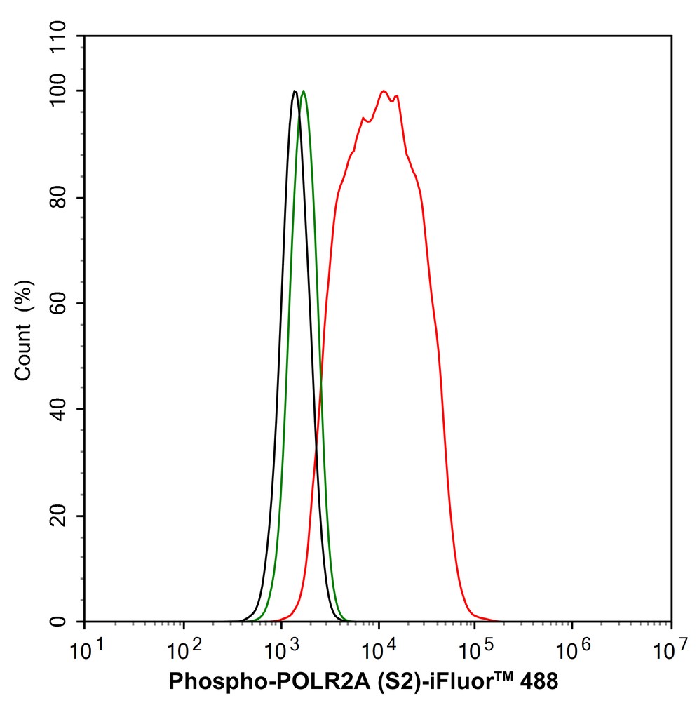
FC
Flow cytometric analysis of Phospho-POLR2A (S2) was done on Hela cells. The cells were fixed, permeabilized and stained with the primary antibody (1/50) (red). After incubation of the primary antibody at room temperature for an hour, the cells were stained with a Alexa Fluor®488 conjugate-Goat anti-Rabbit IgG Secondary antibody at 1/1,000 dilution for 30 minutes.Unlabelled sample was used as a control (cells without incubation with primary antibody; black).| Application Notes | WB:1:500-1:2000 IHC:1:50-1:200 ICC:1:100-1:500 FC:1:50-1:100 |
|---|
| Form | Liquid |
|---|---|
| Storage Instructions | Store at +4℃ after thawing. Aliquot store at -20℃ or -80℃. Avoid repeated freeze / thaw cycles. |
| Storage Buffer | 1*TBS (pH7.4), 0.05% BSA, 40% Glycerol. Preservative: 0.05% Sodium Azide. |
抱歉,暂无相关文献







