| 存储条件 |
|---|
| Product Name | Cathepsin D |
|---|---|
| Antibody Type | Primary Antibodies |
| Antigen Alias | CatD antibody CATD_HUMAN antibody Cathepsin D antibody Cathepsin D heavy chain antibody CD antibody Ceroid lipofuscinosis neuronal 10 antibody CLN10 antibody CPSD antibody ctsd antibody Epididymis secretory sperm binding protein Li 130P antibody HEL S 130P antibody Lysosomal aspartyl peptidase antibody Lysosomal aspartyl protease antibody MGC2311 antibody |
| Product description | The cathepsin family of proteolytic enzymes contains several diverse classes of proteases. The cysteine protease class comprises cathepsins B, L, H, K, S, and O. The aspartyl protease class is composed of cathepsins D and E. Cathepsin G is in the serine protease class. Most cathepsins are lysosomal and each is involved in cellular metabolism, participating in various events such as peptide biosynthesis and protein degradation. Cathepsins may also cleave some protein precursors, thereby releasing regulatory peptides. The promoter region of the cathepsin D gene contains five Sp1 binding sites and four AP-2 binding sites. |
| Immunogen | Recombinant protein |
| Clonality | Polyclonal |
|---|---|
| Isotype | IgG |
| Host Species | Rabbit |
| Tested Applications | WBIHCFC |
| WB:1:500-1:2,000 IHC:1:50-1:200 FC:1:50-1:100 | |
| Species Reactivity | HumanMouseRat |
| Concentration | 1mg/ml |
| Alternative Names | CatD antibody CATD_HUMAN antibody Cathepsin D antibody Cathepsin D heavy chain antibody CD antibody Ceroid lipofuscinosis neuronal 10 antibody CLN10 antibody CPSD antibody ctsd antibody Epididymis secretory sperm binding protein Li 130P antibody HEL S 130P antibody Lysosomal aspartyl peptidase antibody Lysosomal aspartyl protease antibody MGC2311 antibody |
|---|---|
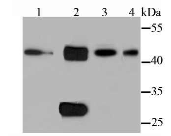
| Molecular Weight(MW) | 45/27 kDa |
| Cellular Localization | Lysosome. Melanosome. Secreted, extracellular space. |
Application
Fig1: Western blot analysis of Cathepsin D on different cell lysates using anti-Cathepsin D antibody at 1/1,000 dilution. Positive control: Lane 1: PC-12 Lane 2: MCF-7 Lane 3: NIH-3T3 Lane 4: Jurkat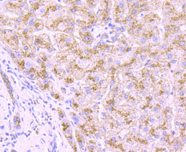
Application
Fig2: Immunohistochemical analysis of paraffin-embedded human liver tissue using anti-Cathepsin D antibody. Counter stained with hematoxylin.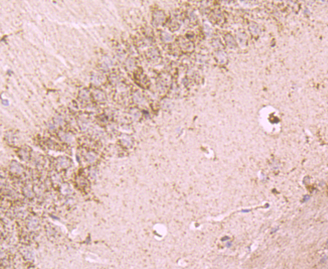
Application
Fig3: Immunohistochemical analysis of paraffin-embedded mouse brain tissue using anti-Cathepsin D antibody. Counter stained with hematoxylin.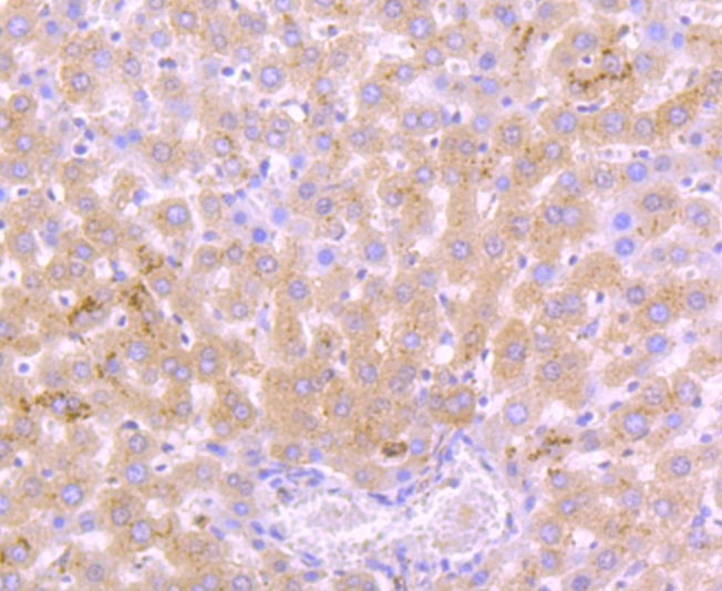
Application
Fig4: Immunohistochemical analysis of paraffin-embedded rat liver tissue using anti-Cathepsin D antibody. Counter stained with hematoxylin.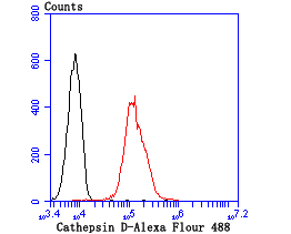
Application
Fig5: Flow cytometric analysis of A549 cells with Cathepsin D antibody at 1/100 dilution (red) compared with an unlabelled control (cells without incubation with primary antibody; black).| Positive Control | PC-12, MCF-7, NIH-3T3, Jurkat, rat liver tissue, human liver tissue, mouse brain tissue. |
|---|---|
| Application Notes | WB:1:500-1:2,000 IHC:1:50-1:200 FC:1:50-1:100 |
| Form | Liquid |
|---|---|
| Storage Instructions | Store at +4℃after thawing. Aliquot store at -20℃or -80℃. Avoid repeated freeze / thaw cycles. |
| Storage Buffer | 1*TBS (pH7.4), 0.5%BSA, 50%Glycerol. Preservative: 0.05% Sodium Azide. |
抱歉,暂无相关文献







