| 存储条件 |
|---|
| Product Name | RON |
|---|---|
| Antibody Type | Primary Antibodies |
| Antigen Alias | c met related tyrosine kinase antibody CD136 antibody CD136 antigen antibodyCDw136 antibody Macrophage stimulating 1 receptor (c met related tyrosine kinase) antibody Macrophage stimulating 1 receptor antibody Macrophage stimulating protein receptor alpha chain antibody MACROPHAGE STIMULATING PROTEIN RECEPTOR antibody Macrophage stimulating protein receptor beta chain antibody Macrophage-Stimulating 1 Receptor (MST1R) antibody Macrophage-stimulating protein receptor beta chain antibody MSP receptor antibody Mst1r antibody MST1R variant RON30 antibody MST1R variant RON62 antibody NPCA3 antibody p185 RON antibody p185-Ron antibody Protein-tyrosine kinase 8 antibody PTK 8 antibody ptk8 antibody PTK8 protein tyrosine kinase 8 antibody Recepteur d’origine nantais (RON) antibody RON antibody RON protein tyrosine kinase antibody RON variant E2E3 antibody RON_HUMAN antibody Soluble RON variant 1 antibody Soluble RON variant 2 antibody Soluble RON variant 3 antibody Soluble RON variant 4 antibody Stem cell derived tyrosine kinase antibody |
| Product description | Receptor tyrosine kinase that transduces signals from the extracellular matrix into the cytoplasm by binding to MST1 ligand. Regulates many physiological processes including cell survival, migration and differentiation. Ligand binding at the cell surface induces autophosphorylation of RON on its intracellular domain that provides docking sites for downstream signaling molecules. Following activation by ligand, interacts with the PI3-kinase subunit PIK3R1, PLCG1 or the adapter GAB1. Recruitment of these downstream effectors by RON leads to the activation of several signaling cascades including the RAS-ERK, PI3 kinase-AKT, or PLCgamma-PKC. RON signaling activates the wound healing response by promoting epithelial cell migration, proliferation as well as survival at the wound site. Plays also a role in the innate immune response by regulating the migration and phagocytic activity of macrophages. Alternatively, RON can also promote signals such as cell migration and proliferation in response to growth factors other than MST1 ligand. |
| Immunogen | Peptide |
| Clonality | Polyclonal |
|---|---|
| Isotype | IgG |
| Host Species | Rabbit |
| Tested Applications | ICCIHC |
| ICC:1:200~1:500 IHC:1:50~1:200 Notes:Optimal dilutions/concentrations should be determined by the researcher. | |
| Species Reactivity | Human |
| Concentration | 1mg/ml |
| Alternative Names | c met related tyrosine kinase antibody CD136 antibody CD136 antigen antibodyCDw136 antibody Macrophage stimulating 1 receptor (c met related tyrosine kinase) antibody Macrophage stimulating 1 receptor antibody Macrophage stimulating protein receptor alpha chain antibody MACROPHAGE STIMULATING PROTEIN RECEPTOR antibody Macrophage stimulating protein receptor beta chain antibody Macrophage-Stimulating 1 Receptor (MST1R) antibody Macrophage-stimulating protein receptor beta chain antibody MSP receptor antibody Mst1r antibody MST1R variant RON30 antibody MST1R variant RON62 antibody NPCA3 antibody p185 RON antibody p185-Ron antibody Protein-tyrosine kinase 8 antibody PTK 8 antibody ptk8 antibody PTK8 protein tyrosine kinase 8 antibody Recepteur d’origine nantais (RON) antibody RON antibody RON protein tyrosine kinase antibody RON variant E2E3 antibody RON_HUMAN antibody Soluble RON variant 1 antibody Soluble RON variant 2 antibody Soluble RON variant 3 antibody Soluble RON variant 4 antibody Stem cell derived tyrosine kinase antibody |
|---|---|
| Molecular Weight(MW) | 152 kDa |
| Cellular Localization | Membrane. |
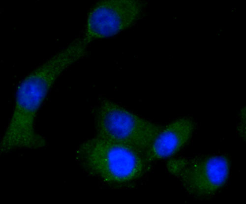
Application
Fig1: ICC staining RON in A549 cells (green). The nuclear counter stain is DAPI (blue). Cells were fixed in paraformaldehyde, permeabilised with 0.25% Triton X100/PBS.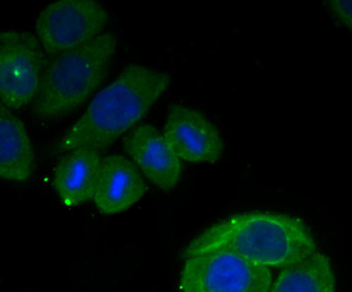
Application
Fig2: ICC staining RON in Ags cells (green). The nuclear counter stain is DAPI (blue). Cells were fixed in paraformaldehyde, permeabilised with 0.25% Triton X100/PBS.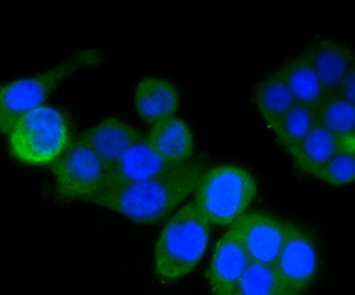
Application
Fig3: ICC staining RON in SW480 cells (green). The nuclear counter stain is DAPI (blue). Cells were fixed in paraformaldehyde, permeabilised with 0.25% Triton X100/PBS.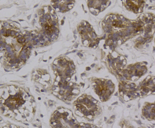
Application
Fig4: Immunohistochemical analysis of paraffin-embedded human breast tissue using anti-RON antibody. Counter stained with hematoxylin.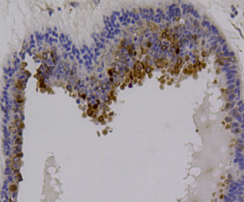
Application
Fig5: Immunohistochemical analysis of paraffin-embedded human breast cancer tissue using anti-RON antibody. Counter stained with hematoxylin.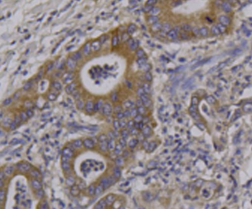
Application
Fig6: Immunohistochemical analysis of paraffin-embedded human stomach cancer tissue using anti-RON antibody. Counter stained with hematoxylin.| Positive Control | A549, Ags, SW480, human breast tissue, human breast cancer tissue, human stomach cancer tissue. |
|---|---|
| Application Notes | ICC:1:200~1:500 IHC:1:50~1:200 Notes:Optimal dilutions/concentrations should be determined by the researcher. |
| Form | Liquid |
|---|---|
| Storage Instructions | Store at +4℃ after thawing. Aliquot store at -20℃ or -80℃. Avoid repeated freeze / thaw cycles. |
| Storage Buffer | 1*TBS (pH7.4), 1%BSA, 40%Glycerol. Preservative: 0.05% Sodium Azide. |
抱歉,暂无相关文献







