| 存储条件 |
|---|
| Product Name | VCAM1 |
|---|---|
| Antibody Type | Primary Antibodies |
| Antigen Alias | CD106 antibody CD106 Antigen antibody INCAM 100 antibody INCAM-100 antibody L1CAM antibody MGC99561 antibody V-CAM 1 antibody Vascular Cell Adhesion Molecule 1 antibody Vascular cell adhesion protein 1 antibody VCAM 1 antibody VCAM-1 antibody VCAM1 antibody VCAM1_HUMAN antibody |
| Product description | Cell adhesion molecules are a family of closely related cell surface glycoproteins involved in cell-cell interactions during growth and are thought to play an important role in embryogenesis and development. Neuronal cell adhesion molecule (NCAM) expression is observed in a variety of human tumors including neuroblastomas, rhabdomyosarcomas, Wilms' tumors, Ewing's sarcomas and some primitive myeloid malignancies. The intracellular adhesion molecule-1 (ICAM-1), also referred to as CD54, is an integral membrane protein of the immunoglobulin superfamily and recognizes the B2α1 and B2αM integrins. PECAM-1 (platelet/endothelial cell adhesion molecule-1), also referred to as CD31, is a glycoprotein expressed on the cell surfaces of monocytes, neutrophils, platelets and a subpopulation of T cells. VCAM-1 (vascular cell adhesion molecule-1) was first identified as an adhesion molecule induced on human endothelial cells by inflammatory cytokines such as IL-1, tumor necrosis factor (TNF) and lipopolysaccharide (LPS). The KALIG gene encodes a nerve cell adhesion molecule (NCAM) -like protein and is deleted in 66% of patients with Kallmann's syndrome, anosmia with secondary hypogonadism. |
| Immunogen | Peptide |
| Clonality | Polyclonal |
|---|---|
| Isotype | IgG |
| Host Species | Rabbit |
| Tested Applications | WBICCIHCFC |
| WB:1:2,000-1:5,000 ICC:1:50-1:200 IHC:1:50-1:200 FC:1:50-1:100 | |
| Species Reactivity | HumanMouseRat |
| Concentration | 1mg/ml |
| Alternative Names | CD106 antibody CD106 Antigen antibody INCAM 100 antibody INCAM-100 antibody L1CAM antibody MGC99561 antibody V-CAM 1 antibody Vascular Cell Adhesion Molecule 1 antibody Vascular cell adhesion protein 1 antibody VCAM 1 antibody VCAM-1 antibody VCAM1 antibody VCAM1_HUMAN antibody |
|---|---|
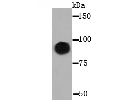
| Molecular Weight(MW) | 100 kDa |
| Cellular Localization | Membrane. |
| SwissProt ID | P19320 |
|---|
Application
Fig1: Western blot analysis of VCAM1 on human spleen tissue lysate using anti-VCAM1 antibody at 1/5,000 dilution.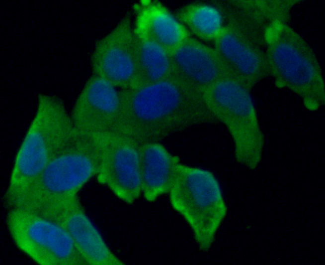
Application
Fig2: ICC staining VCAM1 in Hela cells (green). The nuclear counter stain is DAPI (blue). Cells were fixed in paraformaldehyde, permeabilised with 0.25% Triton X100/PBS.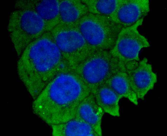
Application
Fig3: ICC staining VCAM1 in JAR cells (green). The nuclear counter stain is DAPI (blue). Cells were fixed in paraformaldehyde, permeabilised with 0.25% Triton X100/PBS.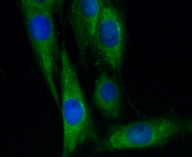
Application
Fig4: ICC staining VCAM1 in SHG-44 cells (green). The nuclear counter stain is DAPI (blue). Cells were fixed in paraformaldehyde, permeabilised with 0.25% Triton X100/PBS.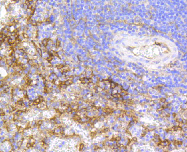
Application
Fig5: Immunohistochemical analysis of paraffin-embedded human spleen tissue using anti-VCAM1 antibody. Counter stained with hematoxylin.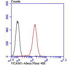
Application
Fig6: Flow cytometric analysis of Hela cells with VCAM1 antibody at 1/100 dilution (red) compared with an unlabelled control (cells without incubation with primary antibody; black).| Positive Control | Human spleen tissue lysate, Hela,JAR, SHG-44, human spleen tissue, HUVEC. |
|---|---|
| Application Notes | WB:1:2,000-1:5,000 ICC:1:50-1:200 IHC:1:50-1:200 FC:1:50-1:100 |
| Form | Liquid |
|---|---|
| Storage Instructions | Store at +4℃after thawing. Aliquot store at -20℃or -80℃. Avoid repeated freeze / thaw cycles. |
| Storage Buffer | 1*TBS (pH7.4), 0.5%BSA, 50%Glycerol. Preservative: 0.05% Sodium Azide. |
抱歉,暂无相关文献







