| 存储条件 |
|---|
| Product Name | CCL3 |
|---|---|
| Antibody Type | Primary Antibodies |
| Antigen Alias | C C motif chemokine 3 antibody CCL 3 antibody CCL3 antibody CCL3_HUMAN antibody Chemokine C C motif ligand 3 antibody G0/G1 switch regulatory protein 19 1 antibody G0/G1 switch regulatory protein 19-1 antibody G0S19 1 antibody G0S19 1 protein antibody Heparin binding chemotaxis protein antibody L2G25B antibody LD78 alpha antibody LD78-alpha(4-69) antibody LD78alpha antibody Macrophage inflammatory protein 1 alpha antibody Macrophage inflammatory protein 1-alpha antibody MIP 1 alpha antibody MIP 1A antibody MIP-1-alpha antibody MIP-1-alpha(4-69) antibody MIP1A antibody PAT 464.1 antibody SCYA 3 antibody SCYA3 antibody SIS alpha antibody SIS beta antibody SIS-beta antibody Small inducible cytokine A3 antibody small inducible cytokine A3 (homologous to mouse Mip-1a) antibody Small-inducible cytokine A3 antibody Tonsillar lymphocyte LD78 alpha protein antibody |
| Product description | Chemokines are members of a superfamily of small inducible, secreted, pro-inflammatory cytokines. Members of the chemokine family exhibit 20 to 50% homology in their predicted amino acid sequences and are divided into four subfamilies. In C-C (or b) subfamily, the first two cysteines are adjacent. C-C chemokines are chemoattractants and activators for monocytes and T cells. C-C subfamily members include macrophage inflammatory protein (MIP)-1α, MIP-1β, MIP-2, MIP-3α, MIP-3β, MIP-4, HCC-1, MIP-5 (or HCC-2), RANTES, MCP-1/2/3 (and the murine homologs JE and MARC), I-309, murine C10 and TCA3. Research has shown that MIP-1β is more selective than MIP-1α, primarily attracting CD4+ T lymphocytes, with a preference for T cells of the naive phenotype. MIP-1α is a more potent lymphocyte chemoattractant than MIP-1β and exhibits a broader range of chemoattractant specificities. It has been suggested that CD8+ T lymphocytes are involved in the control of HIV infection in vivo by the release of HIV-suppressive factors (HIV-SF). MIP-1α has been identified as one of the major HIV-SFs produced by CD8+ T cells, along with MIP-1β and RANTES. |
| Immunogen | peptide |
| Clonality | Polyclonal |
|---|---|
| Isotype | IgG |
| Host Species | Rabbit |
| Tested Applications | ICCIHCFCICC/IF |
| ICC:1:50-1:200 IHC:1:50-1:200 FC:1:50-1:100 WB:1:500 | |
| Species Reactivity | HumanMouse |
| Concentration | 1mg/ml |
| Alternative Names | C C motif chemokine 3 antibody CCL 3 antibody CCL3 antibody CCL3_HUMAN antibody Chemokine C C motif ligand 3 antibody G0/G1 switch regulatory protein 19 1 antibody G0/G1 switch regulatory protein 19-1 antibody G0S19 1 antibody G0S19 1 protein antibody Heparin binding chemotaxis protein antibody L2G25B antibody LD78 alpha antibody LD78-alpha(4-69) antibody LD78alpha antibody Macrophage inflammatory protein 1 alpha antibody Macrophage inflammatory protein 1-alpha antibody MIP 1 alpha antibody MIP 1A antibody MIP-1-alpha antibody MIP-1-alpha(4-69) antibody MIP1A antibody PAT 464.1 antibody SCYA 3 antibody SCYA3 antibody SIS alpha antibody SIS beta antibody SIS-beta antibody Small inducible cytokine A3 antibody small inducible cytokine A3 (homologous to mouse Mip-1a) antibody Small-inducible cytokine A3 antibody Tonsillar lymphocyte LD78 alpha protein antibody |
|---|---|
| Cellular Localization | Secreted |
| SwissProt ID | P10855 |
|---|
Application
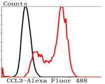
Fig7: Flow cytometric analysis of Hela cells with CCL3 antibody at 1/100 dilution (red) compared with an unlabelled control (cells without incubation with primary antibody; black). Alexa Fluor 488-conjugated Goat anti rabbit IgG was used as the secondary antibody.Application
Fig7: Flow cytometric analysis of Hela cells with CCL3 antibody at 1/100 dilution (red) compared with an unlabelled control (cells without incubation with primary antibody; black). Alexa Fluor 488-conjugated Goat anti rabbit IgG was used as the secondary antibody.Application
Fig7: Flow cytometric analysis of Hela cells with CCL3 antibody at 1/100 dilution (red) compared with an unlabelled control (cells without incubation with primary antibody; black). Alexa Fluor 488-conjugated Goat anti rabbit IgG was used as the secondary antibody.Application
Fig7: Flow cytometric analysis of Hela cells with CCL3 antibody at 1/100 dilution (red) compared with an unlabelled control (cells without incubation with primary antibody; black). Alexa Fluor 488-conjugated Goat anti rabbit IgG was used as the secondary antibody.Application
Fig7: Flow cytometric analysis of Hela cells with CCL3 antibody at 1/100 dilution (red) compared with an unlabelled control (cells without incubation with primary antibody; black). Alexa Fluor 488-conjugated Goat anti rabbit IgG was used as the secondary antibody.Application
Fig7: Flow cytometric analysis of Hela cells with CCL3 antibody at 1/100 dilution (red) compared with an unlabelled control (cells without incubation with primary antibody; black). Alexa Fluor 488-conjugated Goat anti rabbit IgG was used as the secondary antibody.Application
Fig7: Flow cytometric analysis of Hela cells with CCL3 antibody at 1/100 dilution (red) compared with an unlabelled control (cells without incubation with primary antibody; black). Alexa Fluor 488-conjugated Goat anti rabbit IgG was used as the secondary antibody.| Positive Control | Hela, PANC-1, A549, mouse pancreas tissue, mouse lung tissue, mouse spleen tissue |
|---|---|
| Application Notes | ICC:1:50-1:200 IHC:1:50-1:200 FC:1:50-1:100 WB:1:500 |
| Form | Liquid |
|---|---|
| Storage Instructions | Store at +4℃ after thawing. Aliquot store at -20℃ or -80℃. Avoid repeated freeze / thaw cycles. |
| Storage Buffer | 1*TBS (pH7.4), 1%BSA, 40%Glycerol. Preservative: 0.05% Sodium Azide. |
抱歉,暂无相关文献







