| 存储条件 |
|---|
| Product Name | Cytochrome C [10-E11-G2] |
|---|---|
| Antibody Type | Primary Antibodies |
| Antigen Alias | CYC antibody CYC_HUMAN antibody CYCS antibody Cytochrome c antibody Cytochrome c somatic antibody HCS antibody THC4 antibody |
| Product description | Cytochrome c is a well characterized mobile electron transport protein that is essential to energy conversion in all aerobic organisms. In mammalian cells, this highly conserved protein is normally localized to the mitochondrial intermembrane space. More recent studies have identifed cytosolic cytochrome c as a factor necessary for activation of apoptosis. During apoptosis, cytochrome c is translocated from the mitochondrial membrane to the cytosol, where it is required for activation of caspase-3 (CPP32). Overexpression of Bcl-2 has been shown to prevent the translocation of cytochrome c, thereby blocking the apoptotic process. Overexpression of Bax has been shown to induce the release of cytochrome c and to induce cell death. The release of cytochrome c from the mitochondria is thought to trigger an apoptotic cascade, whereby Apaf-1 binds to Apaf-3 (caspase-9) in a cytochrome c-dependent manner, leading to caspase-9 cleavage of caspase-3. |
| Immunogen | Peptide |
| Clonality | Monoclonal |
|---|---|
| Isotype | IgG1 |
| Host Species | Mouse |
| Tested Applications | WBIHCICC |
| WB:1:250~1:1000 IHC:1:20~1:100 ICC:1:100~1:200 Notes:Optimal dilutions/concentrations should be determined by the researcher. | |
| Species Reactivity | HumanMouseRat |
| Concentration | 2mg/ml |
| Alternative Names | CYC antibody CYC_HUMAN antibody CYCS antibody Cytochrome c antibody Cytochrome c somatic antibody HCS antibody THC4 antibody |
|---|---|
| Molecular Weight(MW) | 12 kDa |
| Cellular Localization | Mitochondrion. |
Application
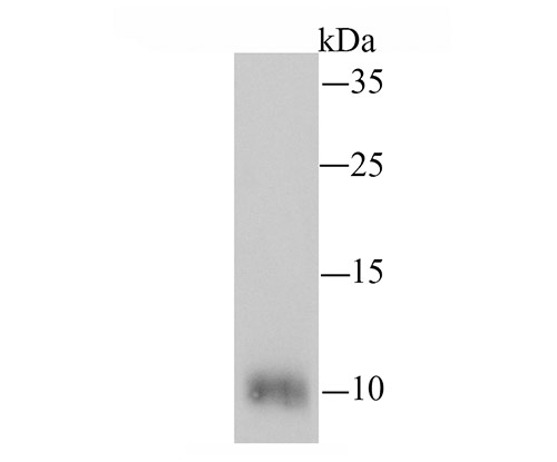
Fig1: Western blot analysis of Cytochrome C on mouse heart tissue lysate using anti-Cytochrome C antibody at 1/200 dilution.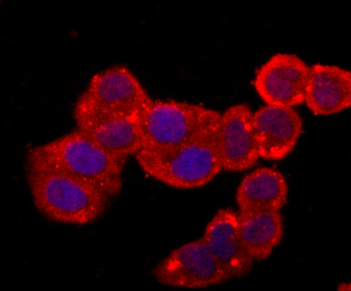
Application
Fig2: ICC staining Cytochrome C (red) in Hela cells. The nuclear counter stain is DAPI (blue). Cells were fixed in paraformaldehyde, permeabilised with 0.25% Triton X100/PBS.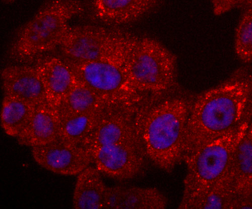
Application
Fig3: ICC staining Cytochrome C (red) in HepG2 cells. The nuclear counter stain is DAPI (blue). Cells were fixed in paraformaldehyde, permeabilised with 0.25% Triton X100/PBS.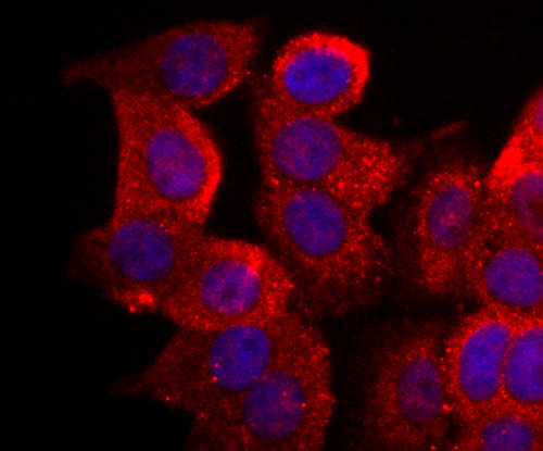
Application
Fig4: ICC staining Cytochrome C (red) in MCF-7 cells. The nuclear counter stain is DAPI (blue). Cells were fixed in paraformaldehyde, permeabilised with 0.25% Triton X100/PBS.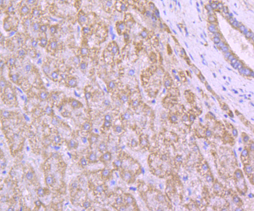
Application
Fig5: Immunohistochemical analysis of paraffin-embedded human liver tissue using anti-Cytochrome C antibody. Counter stained with hematoxylin.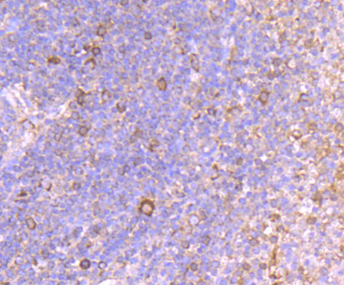
Application
Fig6: Immunohistochemical analysis of paraffin-embedded human spleen tissue using anti-Cytochrome C antibody. Counter stained with hematoxylin.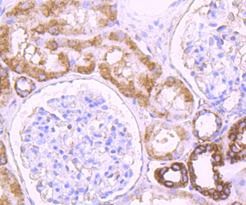
Application
Fig7: Immunohistochemical analysis of paraffin-embedded human kidney tissue using anti-Cytochrome C antibody. Counter stained with hematoxylin.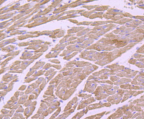
Application
Fig8: Immunohistochemical analysis of paraffin-embedded mouse heart tissue using anti-Cytochrome C antibody. Counter stained with hematoxylin.| Positive Control | Mouse heart tissue lysate, Hela, HepG2, MCF-7, human liver tissue, human spleen tissue, human kidney tissue, mouse heart tissue. |
|---|---|
| Application Notes | WB:1:250~1:1000 IHC:1:20~1:100 ICC:1:100~1:200 Notes:Optimal dilutions/concentrations should be determined by the researcher. |
| Form | Liquid |
|---|---|
| Storage Instructions | Store at +4℃ after thawing. Aliquot store at -20℃ or -80℃. Avoid repeated freeze / thaw cycles. |
| Storage Buffer | 1*TBS (pH7.4), 0.5%BSA, 50%Glycerol. Preservative: 0.05% Sodium Azide. |
抱歉,暂无相关文献







