Anti-Oligodendrocyte Specific蛋白抗体- Oligodendrocyte Marker
参阅全部 Oligodendrocyte Specific Protein 一抗
兔多克隆抗体to Oligodendrocyte Specific蛋白- Oligodendrocyte Marker
Rabbit
Reacts specifically with 22 kDa protein derived from CNS samples.
适用于: IHC-P, IHC-Frmore details
与反应: Rat, Human
A 15 residue synthetic C-terminal peptide.
The concentration depends on the batch, please enquire for current stock concentration.
The Life Science industry has been in the grips of a reproducibility crisis for a number of years. Abcam is leading the way in addressing this with our range of recombinant monoclonal antibodies and knockout edited cell lines for gold-standard validation. Please check that this product meets your needs before purchasing.
If you have any questions, special requirements or concerns, please send us an inquiry and/or contact our Support team ahead of purchase. Recommended alternatives for this product can be found below, along with publications, customer reviews and Q&As
Liquid
Shipped at 4°C. Store at +4°C short term (1-2 weeks). Upon delivery aliquot. Store at -20°C or -80°C. Avoid freeze / thaw cycle.
Constituent: Whole serum
Whole antiserum
多克隆
IgG
Abpromise™承诺保证使用ab7474于以下的经测试应用
“应用说明”部分 下显示的仅为推荐的起始稀释度;实际最佳的稀释度/浓度应由使用者检定。
| 应用 | Ab评论 | 说明 |
|---|---|---|
| IHC-P | (2) | 1/50 - 1/500. |
| IHC-Fr | (1) | Use at an assay dependent concentration. |
Entrez Gene: 5010 Human
Omim: 601326 Human
SwissProt: O75508 Human
Unigene: 31595 Human
Unigene: 8282 Rat
Cldn11 antibody
Oligodendrocyte transmembrane protein antibody
Oligodendrocyte-specific protein antibody
OSP antibody
OTM antibody
Claudin 11 antibody
Claudin-11 antibody
CLD11_HUMAN antibody

Immunohistochemistry (Frozen sections) - Anti-Oligodendrocyte Specific Protein antibody - Oligodendrocyte Marker (ab7474)This image is courtesy of an Abreview submitted by Dr Qin Wen
Immunohistochemical analysis of adult mouse testis tissue, staining Oligodendrocyte Specific Protein (red) with ab7474.
Tissue was blocked with 5% BSA for 1 hour at 25°C. Samples were incubated with primary antibody (1/100 in 1% BSA) for 1 hour at 25°C. A TRITC-conjugated donkey anti-rabbit polyclonal IgG (1/200) was used as the secondary antibody.
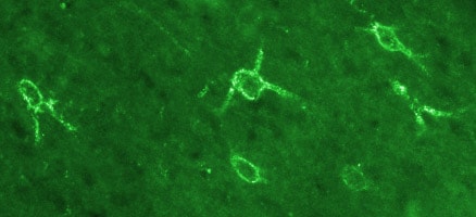
Immunohistochemistry (Frozen sections) - Anti-Oligodendrocyte Specific Protein antibody - Oligodendrocyte Marker (ab7474)
Immunofluorescent staining for OSP obtained with OSP antibody (ab7474) in rat brain cortex. The picture shows oligodendrocyte immunostaining in the cingular cortex. Picture taken with X20 objective, cells stained are ~30microns in diameter. Animals were intracardially perfused with 4% PFA. Tissue was post-fixed overnight in the same fixative, cryoprotected in 20% sucrose and frozen in OCT. Protocol : free floating IHC on 30um cryostat sections. Primary antibody ab7474 was used at 1/100 and incubated overnight at RT. Secondary antibody, Alexa fluor 488 at 1/1000, incubated for 2h at RT.
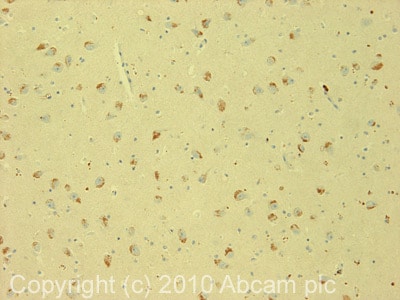
Immunohistochemistry (Formalin/PFA-fixed paraffin-embedded sections) - Anti-Oligodendrocyte Specific Protein antibody - Oligodendrocyte Marker (ab7474)
IHC image of ab7474 staining in human hippocampus formalin fixed paraffin embedded tissue section, performed on a Leica BondTM system using the standard protocol F. The section was pre-treated using heat mediated antigen retrieval with sodium citrate buffer (pH6, epitope retrieval solution 1) for 20 mins. The section was then incubated with ab7474, 5µg/ml, for 15 mins at room temperature and detected using an HRP conjugated compact polymer system. DAB was used as the chromogen. The section was then counterstained with haematoxylin and mounted with DPX.
For other IHC staining systems (automated and non-automated) customers should optimize variable parameters such as antigen retrieval conditions, primary antibody concentration and antibody incubation times.


