山羊抗兔IgG H&L (HRP)
参阅全部 IgG 二抗
Goat
Rabbit
适用于: IHC-P, WB, ELISA, Immunomicroscopy, Dot blot, ICC, IHC-Frmore details
Rabbit IgG, whole molecule
HRP
Liquid
Shipped at 4°C. Store at +4°C short term (1-2 weeks). Upon delivery aliquot. Store at -20°C. Avoid freeze / thaw cycle. Please see notes section. Store In the Dark.
Preservative: 0.01% Gentamicin sulphate
Constituents: 1% BSA, 0.424% Potassium phosphate solution, 0.88% Sodium chloride
Immunogen affinity purified
RABBIT IgG (H&L) Secondary Antibody Peroxidase Conjugated was prepared from monospecific antiserum by immunoaffinity chromatography using Rabbit IgG coupled to agarose beads.
Horseradish Peroxidase (HRP)
多克隆
IgG
Abpromise™承诺保证使用ab6721于以下的经测试应用
“应用说明”部分 下显示的仅为推荐的起始稀释度;实际最佳的稀释度/浓度应由使用者检定。
| 应用 | Ab评论 | 说明 |
|---|---|---|
| IHC-P | (2) | 1/1000. |
| WB | (10) | 1/2000 - 1/20000. Suggested working dilution of 1/3000 (see PMID: 17222046). In addition, found to work at 1/20000 (see PMID: 16936283). Working dilutions are highlighted in the table below. Please note that the antibody can be diluted to 1:48,000 to 1:207,000 in many instances. Read More |
| ELISA | 1/120000. | |
| Immunomicroscopy | Use at an assay dependent concentration. | |
| Dot blot | Use at an assay dependent concentration. | |
| ICC | 1/1000 - 1/5000. | |
| IHC-Fr | 1/1000. |

Immunohistochemistry (Formalin/PFA-fixed paraffin-embedded sections) - Goat Anti-Rabbit IgG H&L (HRP) (ab6721)Farah et al PLoS One. 2018 Feb 2;13(2):e0191526. doi: 10.1371/journal.pone.0191526. eCollection 2018. Fig 3. Reproduced under the Creative Commons license http://creativecommons.org/licenses/by/4.0/
Anti-phospho-tau immunostaining in Dryocopus lineatus.
Tau-positivity in the midbrain (Panel A (shown) and B) and the corpus callosum (C) of the Dryocopus lineatus brain. The axonal tract staining demonstrates a thread-like pattern, similar to that seen with Gallyas sliver staining. Occasional intracellular tau-accumulations were identified within neurons (D).
For full method, please see paper.
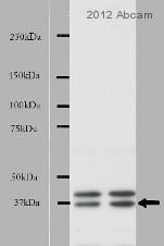
Western blot - Goat Anti-Rabbit IgG H&L (HRP) (ab6721)This image is courtesy of an Abreview submitted by Maryna Polyakova.
All lanes : Anti-NEK7 antibody (ab80948) at 1/2000 dilution
All lanes : Mouse brain tissue cytoplasmic lysate
Lysates/proteins at 10 µg per lane.
Secondary
All lanes : Goat Anti-Rabbit IgG H&L (HRP) (ab6721) at 1/5000 dilution
Developed using the ECL technique.
Performed under reducing conditions.
Exposure time: 10 seconds
Blocked with 5% non-fat milk for 1 hour at 18°C

Immunohistochemistry (Formalin/PFA-fixed paraffin-embedded sections) - Goat Anti-Rabbit IgG H&L (HRP) (ab6721)This image is courtesy of an anonymous Abreview.
ab3580 staining glucocorticoid receptor in mouse epididymis tissue sections by Immunohistochemistry (IHC-P - paraformaldehyde-fixed, paraffin-embedded sections).
Tissue was fixed with Bouin's solution and blocked with 1.5% serum for 30 minutes at 25°C; antigen retrieval was by heat mediation in a citrate buffer. Samples were incubated with primary antibody (1/1000) for 14 hours at 4°C. An HRP-conjugated goat anti-rabbit IgG H&L (ab6721) (1/200) was used as the secondary antibody.

Immunohistochemistry (Formalin/PFA-fixed paraffin-embedded sections) - Goat Anti-Rabbit IgG H&L (HRP) (ab6721)Image is courtesy of an anonymous AbReview.
Immunohistochemical analysis of PFA-fixed paraffin-embedded rat cardiac tissue sections, labeling Conexin 43 with ab117843 at a dilution of 1/500 incubated for 12 hours at 4°C in 1% BSA in TBS. Antigen retrival was via Tris-EDTA pH 9.0 (heat mediated). Blocking was 3% BSA incubated for 1 hour at 37°C. The secondary was ab6721 at 1/500.

Immunohistochemistry (Formalin/PFA-fixed paraffin-embedded sections) - Goat Anti-Rabbit IgG H&L (HRP) (ab6721)
ab6721 was used at dilution 1/100 with the primary antibody ab11370 in IHC-P. See the review on ab11370.

Immunohistochemistry (Frozen sections) - Goat Anti-Rabbit IgG H&L (HRP) (ab6721)
ab6721 was used at dilution 1/100 with the primary antibody ab35604 in IHC-Fr. See the review on ab35604.


