山羊抗兔IgG H&L (Cy5 ®)预吸附二抗
参阅全部 IgG 二抗
Goat
Rabbit
适用于: Flow Cyt, ICC/IF, ELISA, IHC-P, IHC-Frmore details
Chicken, Cow, Goat, Guinea pig, Hamster, Horse, Human, Mouse, Rat, Sheepmore details
Rabbit IgG, whole molecule
Cy5 ®. Ex: 650nm, Em: 667nm
Liquid
Shipped at 4°C. Store at +4°C.
pH: 6.50
Preservative: 0.01% Sodium azide
Constituents: 0.42% Potassium phosphate, 0.87% Sodium chloride, 1% BSA
浓度
500 µg 浓度为 1 mg/ml
Immunogen affinity purified
This product was prepared from monospecific antiserum by immunoaffinity chromatography using Rabbit IgG coupled to agarose beads followed by solid phase adsorption(s) to remove any unwanted reactivities and extensive dialysis.
Cy5.29 (Cyanine 5.29-OSu) (Molecular Weight 975 daltons) Absorption Wavelength: 650 nm Emission Wavelength: 667 nm Fluorochrome/Protein Ratio: 6.0 moles Cy5 per mole of Goat IgG
多克隆
IgG
Cy™ and CyDye™ are registered trademarks of Cytiva.
This secondary antibody is specifically designed for the detection of multiple primary antibodies (polyclonal or monoclonal) of different host species in experiments where cells are simultaneously labeled without unwanted cross reaction.
Abpromise™承诺保证使用ab6564于以下的经测试应用
“应用说明”部分 下显示的仅为推荐的起始稀释度;实际最佳的稀释度/浓度应由使用者检定。
| 应用 | Ab评论 | 说明 |
|---|---|---|
| Flow Cyt | (1) | Use at an assay dependent dilution. |
| ICC/IF | 1/1000 - 1/5000. | |
| ELISA | 1/10000 - 1/50000. | |
| IHC-P | Use at an assay dependent concentration. | |
| IHC-Fr | Use at an assay dependent concentration. |

Immunohistochemistry (Frozen sections) - Goat Anti-Rabbit IgG H&L (Cy5 ®) preadsorbed (ab6564)Hennenberg, M. et al PLoS One. 2012;7(11):e50904. doi: 10.1371/journal.pone.0050904. Epub 2012 Nov 30 Reproduced under the Creative Commons license http://creativecommons.org/licenses/by/4.0/
Immunofluorescence stainings of human prostate tissues
Sections were double labeled for different PAK isoforms, together with calponin (marker for smooth muscle cells), pan-cytokeratin (marker for epithelial cells of glands), or tyrosine hydroxylase (TH, marker for catecholaminergic nerves). Yellow color in merged pictures may indicate colocalization of targets. Shown are representative stainings from series with tissues from n = 5 patients for each combination.
ab6908 was used as secondary antibody to visualize various rabbit anti-PAK primary antibodies (red).
(From Figure 2 of Hennenberg et al)

Immunohistochemistry (Formalin/PFA-fixed paraffin-embedded sections) - Goat Anti-Rabbit IgG H&L (Cy5 ®) preadsorbed (ab6564)This image is courtesy of an anonymous Abreview.
ab11175 staining Histone H2A.X in mouse testis tissue sections by Immunohistochemistry (IHC-P - paraformaldehyde-fixed, paraffin-embedded sections). Tissue was fixed with paraformaldehyde, permeabilized with 0.1% Triton X-100, and blocked with 4% BSA for 2 hours at 37°C. Antigen retrieval was by heat mediation in a citrate buffer. Samples were incubated with primary antibody (1/100 in PBS/4% BSA/0.05% Triton X-100) for 48 hours at 4°C. A Cy5®-conjugated goat anti-rabbit IgG H&L preadsorbed (ab6564) (1/300) was used as the secondary antibody.

Flow Cytometry - Goat Anti-Rabbit IgG H&L (Cy5 ®) preadsorbed (ab6564)
ab6564 was used at dilution 1/300 with the primary antibody ab19898 in Flow Cyt. See the review on ab19898.
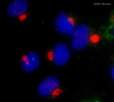
Immunocytochemistry/ Immunofluorescence - Goat Anti-Rabbit IgG H&L (Cy5 ®) preadsorbed (ab6564)
ab6564 was used at dilution 1/500 with the primary antibody ab24586 in ICC/IF. See the review on ab24586.


