Anti-alpha smooth muscle Actin抗体
参阅全部 alpha smooth muscle Actin 一抗
兔多克隆抗体to alpha smooth muscle Actin
Rabbit
Alpha smooth muscle actin antibody (ab5694) stains smooth muscle cells in vessel walls, gut wall, and myometrium. Myoepithelial cells in breast and salivary gland are also stained. ab5694 reacts with tumors arising from smooth muscles and myoepithelial cells.
适用于: WB, IHC-Pmore details
与反应: Mouse, Human
Synthetic peptide corresponding to Human alpha smooth muscle Actin (N terminal).
Database link: P62736
WB: HEK-293, A431, HeLa, Jurkat and NIH/3T3 whole cell lysate. Mouse heart tissue homogenate. HeLa nuclear lysate. IHC-P: PACT-sRIMS-cleared virgin and lactating mammary glands. Mouse intestine and pancreas tissue. Mouse aortic regurgitation. Mouse tonsil tissue.
Actins are highly conserved proteins expressed in all eucaryotic cells. Actin filaments form part of the cytoskeleton and play essential roles in regulating cell shape and movement. Six distinct actin isotypes have been identified in mammalian cells. Each is encoded by a separated gene and is expressed in a developmentally regulated and tissue-specific manner, alpha and beta cytoplasmic actins are expressed in a wide variety of cells; whereas, expression of alpha skeletal, alpha cardiac, alpha vascular and gamma enteric actins are more restricted to specialized muscle cell type. Smooth muscle alpha actin is of further interest because it is one of a few genes whose expression is relatively restricted to vascular smooth muscle cells. Further more, expression of smooth muscle alpha actin is regulated by hormones, cell proliferation and altered by pathological conditions including oncogenic transformation and atherosclerosis.
Abcam is committed to meeting high standards of manufacturing and has decided to discontinue this product once the stock runs out as we are unable to secure its future high-quality supply. We suggest ab124964, ab150301 or ab7817 as possible replacements. We are sorry for any inconvenience this may cause.
The Life Science industry has been in the grips of a reproducibility crisis for a number of years. Abcam is leading the way in addressing this with our range of recombinant monoclonal antibodies and knockout edited cell lines for gold-standard validation. Please check that this product meets your needs before purchasing.
If you have any questions, special requirements or concerns, please send us an inquiry and/or contact our Support team ahead of purchase. Recommended alternatives for this product can be found below, along with publications, customer reviews and Q&As
Liquid
Shipped at 4°C. Store at +4°C short term (1-2 weeks). Upon delivery aliquot. Store at -20°C long term. Avoid freeze / thaw cycle.
pH: 7.40
Preservative: 0.05% Sodium azide
Constituent: 99.95% PBS
浓度
100 µg 浓度为 0.2 mg/ml
Immunogen affinity purified
多克隆
IgG
Abpromise™承诺保证使用ab5694于以下的经测试应用
“应用说明”部分 下显示的仅为推荐的起始稀释度;实际最佳的稀释度/浓度应由使用者检定。
| 应用 | Ab评论 | 说明 |
|---|---|---|
| WB | (28) | Use a concentration of 0.5 - 2 µg/ml. Predicted molecular weight: 42 kDa. |
| IHC-P | (69) | 1/50 - 1/200. Perform heat mediated antigen retrieval before commencing with IHC staining protocol. |
Entrez Gene: 59 Human
Entrez Gene: 11475 Mouse
Omim: 102620 Human
SwissProt: P62736 Human
SwissProt: P62737 Mouse
Unigene: 500483 Human
Unigene: 213025 Mouse
ACTA2 antibody
Actin alpha 2 smooth muscle aorta antibody
Actin aortic smooth muscle antibody
Actin, aortic smooth muscle antibody
ACTSA antibody
ACTVS antibody
Alpha 2 actin antibody
Alpha actin 2 antibody
Alpha cardiac actin antibody
alpha sma antibody
Alpha-actin-2 antibody
Cell growth inhibiting gene 46 protein antibody
Cell growth-inhibiting gene 46 protein antibody
GIG46 antibody
Growth inhibiting gene 46 antibody
MYMY5 antibody
a actin antibody
AAT6 antibody
ACTA_HUMAN antibody

Immunohistochemistry (Formalin/PFA-fixed paraffin-embedded sections) - Anti-alpha smooth muscle Actin antibody (ab5694)This image is courtesy of an Abreview submitted by Cameron Johnstone.
Immunohistochemistry (Formalin/PFA-fixed paraffin-embedded sections) analysis of 10% formalin-fixed Human transitional cell carcinoma of the kidney. Stained with ab5694 at 1/150 dilution. Secondary antibody used was goat anti-rabbit Alexa-568® at 1/500 dilution. Blocking was done with 5% serum for 1 hour at 25°C. The sample was incubated with the primary antibody for 1hour at 25°C with 5% normal goat serum. Antigen retrieval method was heat mediated with EDTA pH 8.

Western blot - Anti-alpha smooth muscle Actin antibody (ab5694)
Lanes 1-3 : Anti-alpha smooth muscle Actin antibody (ab5694) at 1 µg/ml
Lane 1 : HEK-293 (Human epithelial cell line from embryonic kidney) cell lysate - overexpressing alpha-Actin
Lane 2 : NIH/3T3 (Mouse embryonic fibroblast cell line) cell lysate
Lanes 3 & 5 : Mouse heart tissue homogenate
Lane 4 : NIH/3T3 cell lysate
Lysates/proteins at 20 µg per lane.
Secondary
All lanes : Fluor 750-conjugated goat anti-rabbit IgG (H+L) at 1/12500 dilution
Predicted band size: 42 kDa
Observed band size: 42 kDa
Incubated with the primary antibody at 4°C overnight.
Incubated with the secondary antibody at room temperature for 1 hour.

Immunohistochemistry (Formalin/PFA-fixed paraffin-embedded sections) - Anti-alpha smooth muscle Actin antibody (ab5694)Image from Lloyd-Lewis B. et al., Breast Cancer Res. 2016 18: 127. Fig 2.; doi: 10.1186/s13058-016-0754-9. Reproduced under the Creative Commons license http://creativecommons.org/licenses/by/4.0/.
Passive CLARITY technique (PACT)-sorbitol refractive index matching solution (sRIMS) clearing and 3D imaging of virgin and lactating mouse mammary tissue. a PACT-sRIMS tissue clearing and immunostaining protocol and timeline. Three-dimensional confocal imaging of PACT-sRIMS-cleared virgin (b) and lactating (c) mammary glands immunostained with basal cell markers (K5 and smooth muscle actin (stained with ab5694)) and luminal cell markers (K8 and E-cadherin (E-CAD)). Main image shows the maximum intensity projection of the entire image sequence, with thin optical slices (1 μm) and their depth (z value) relative to the first image in the image sequence.
From a paper comparing imaging of intact virgin and lactating mammary glands using 3D imaging of solvent-cleared organs, see deep brain (seeDB), clear unobstructed brain imaging cocktails (CUBIC) and passive clarity technique.

Immunohistochemistry (Formalin/PFA-fixed paraffin-embedded sections) - Anti-alpha smooth muscle Actin antibody (ab5694)
This picture shows formalin-fixed, paraffin embedded mouse intestine and mesentery, the optimal dilution is 1:1600 to 1:3200, incubation overnight at 4oC, counterstained with Hematoxylin.
This image was kindly supplied as part of the review by JQ Zhang.

Immunohistochemistry (Formalin/PFA-fixed paraffin-embedded sections) - Anti-alpha smooth muscle Actin antibody (ab5694)Image from Lee H. et al., BMC Dev Biol. 2014 Dec 21;14:48. Fig 2Bdoi: 10.1186/s12861-014-0048-3 Reproduced under the Creative Commons license http://creativecommons.org/licenses/by/4.0/.
Pancreatic vessel imaging in the intact adult mouse pancreas. In adult mouse tissues (12 weeks old), imaging was performed after CLARITY. Three-dimensional projection clarified mouse pancreas with capillary immunostained for α-smooth muscle actin (green). Scale bar, 200 μm.
From a study, that aimed to improve the original CLARITY procedure and developed specific CLARITY protocols for various intact organs.
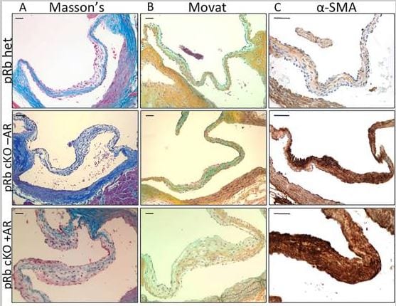
Immunohistochemistry (Formalin/PFA-fixed paraffin-embedded sections) - Anti-alpha smooth muscle Actin antibody (ab5694)Freytsis, M. et al PLoS One. 2018 Jan 5;13(1):e0190623. doi: 10.1371/journal.pone.0190623. eCollection 2018 Reproduced under the Creative Commons license http://creativecommons.org/licenses/by/4.0/
Representative histology of aortic valve leaflets from aged mice demonstrates changes in pRb cKO AoV
A) Masson’s trichrome showing reduced collagen staining (blue) in leaflet from pRb cKO mouse with aortic regurgitation (AR). B) Movat pentachrome showing more diffuse collage staining (yellow) in fibrosa, but normal proteoglycan staining (blue) in the spongiosa layer of the leaflet from pRb cKO with AR. C) Immunohistochemistry for α-SMA, demonstrating presence of activated myofibroblasts throughout leaflets of pRb cKO mouse with and without AR. Scale bar is 50μm.
Alpha smooth muscle Actin is detected witn ab5694 at 1/1000 dilution.
(From Figure 2 of Freytsis et al)

Immunohistochemistry (Formalin/PFA-fixed paraffin-embedded sections) - Anti-alpha smooth muscle Actin antibody (ab5694)
Immunohistochemistry (Formalin/PFA-fixed paraffin-embedded sections) analysis of human tonsil tissue labelling alpha smooth muscle Actin with ab5694 at a dilution of 1/1000. Heat mediated antigen retrieval was performed for 35 minutes followed by cooling for 20 minutes. Sections were incubated with the primary antibody for 1 hour followed by incubation with a biotinylated secondary antibody for 30 minutes then HRP-Streptavidin for 30 minutes. Developed using DAB chromogen substrate (5-10 minutes). Counter stained with hematoxylin.
Magnification: left - 10X, right - 40X.
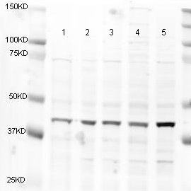
Western blot - Anti-alpha smooth muscle Actin antibody (ab5694)
All lanes : Anti-alpha smooth muscle Actin antibody (ab5694) at 1 µg/ml
Lane 1 : HeLa (Human epithelial cell line from cervix adenocarcinoma) nuclear cell lysate
Lane 2 : HeLa whole cell lysate
Lane 3 : A431 (Human epidermoid carcinoma cell line) cell lysate
Lane 4 : Jurkat (Human T cell leukemia cell line from peripheral blood) cell lysate
Lane 5 : HEK-293 (Human epithelial cell line from embryonic kidney) cell lysate
Lysates/proteins at 20 µg per lane.
Secondary
All lanes : Alexa Fluor anti-rabbit at 1/5000 dilution
Performed under reducing conditions.
Predicted band size: 42 kDa
Observed band size: 42 kDa
Additional bands at: 30 kDa, 35 kDa, 37 kDa, 50 kDa, 75 kDa. We are unsure as to the identity of these extra bands.
Please note that ab5694 does not appear to be specific to smooth muscle.


