Anti-Histone H3 (citrulline R2 + R8 + R17)抗体
参阅全部 Histone H3 一抗
兔多克隆抗体to Histone H3 (citrulline R2 + R8 + R17)
Rabbit
ab5103 detects a 17 kDa band in single lane Western Blot. Peptide inhibition in Western Blot hasn't been processed. Modification specificity is determined by Peptide Array. ab5103 binds strongly to Histone H3 citrulline 2 + 8 + 17 peptide.
From Mar 2024, QC testing of replenishment batches of this polyclonal changed. All tested and expected application and reactive species combinations are still covered by our Abcam product promise. However, we no longer test all applications. For more information on a specific batch, please contact our Scientific Support who will be happy to help. You may also be interested in our alternative recombinant antibody, ab281584.
适用于: PepArr, IP, WBmore details
不适用于: ICC/IF
与反应: Mouse, Rat, Human, Recombinant fragment
预测可用于: Rabbit, Cow, Monkey, a wide range of other species![]()
Synthetic peptide. This information is proprietary to Abcam and/or its suppliers.
WB: Mouse and rat brain tissue lysates. HL60 + DMSO, NIH/3T3 and PC12 whole cell lysates. IP: HEK-293T (human embryonic kidney) transfected with PADI4 expression vector containing a GFP-tag
The Life Science industry has been in the grips of a reproducibility crisis for a number of years. Abcam is leading the way in addressing this with our range of recombinant monoclonal antibodies and knockout edited cell lines for gold-standard validation. Please check that this product meets your needs before purchasing.
If you have any questions, special requirements or concerns, please send us an inquiry and/or contact our Support team ahead of purchase. Recommended alternatives for this product can be found below, along with publications, customer reviews and Q&As
Liquid
Shipped at 4°C. Store at +4°C short term (1-2 weeks). Upon delivery aliquot. Store at -20°C or -80°C. Avoid freeze / thaw cycle.
pH: 7.40
Preservative: 0.02% Sodium azide
Constituents: 98.98% PBS, 1% BSA
Batches of this product that have a concentration < 1mg/ml may have BSA added as a stabilising agent. If you would like information about the formulation of a specific lot, please contact our scientific support team who will be happy to help.
Immunogen affinity purified
多克隆
IgG
Abpromise™承诺保证使用ab5103于以下的经测试应用
“应用说明”部分 下显示的仅为推荐的起始稀释度;实际最佳的稀释度/浓度应由使用者检定。
| 应用 | Ab评论 | 说明 |
|---|---|---|
| PepArr | Use a concentration of 0.2 - 0.02 µg/ml. | |
| IP | Use at an assay dependent concentration. | |
| WB | (9) | Use a concentration of 1 µg/ml. Detects a band of approximately 17 kDa (predicted molecular weight: 15 kDa). In our hands, when tested in western blot, this product typically gives a weaker signal in mouse and rat tissue lysates compared to mouse and rat cell lines. Abcam welcomes customer feedback and would appreciate any comments regarding this product and the data presented above. Abcam recommends using 3-5% milk as the blocking agent We recommend Goat Anti-Rabbit IgG H&L (HRP) (ab97051) secondary antibody. Read More |
应用说明
Is unsuitable for ICC/IF.
Entrez Gene: 8350 Human
Entrez Gene: 8351 Human
Entrez Gene: 8352 Human
Entrez Gene: 8353 Human
Entrez Gene: 8354 Human
Entrez Gene: 8355 Human
Entrez Gene: 8356 Human
Entrez Gene: 8357 Human
Entrez Gene: 8358 Human
Entrez Gene: 8968 Human
Entrez Gene: 319152 Mouse
Entrez Gene: 319153 Mouse
Entrez Gene: 360198 Mouse
Entrez Gene: 97908 Mouse
Omim: 602810 Human
SwissProt: P68431 Human
SwissProt: P68433 Mouse
Unigene: 132854 Human
Unigene: 247813 Human
Unigene: 247814 Human
Unigene: 248176 Human
Unigene: 443021 Human
Unigene: 484990 Human
Unigene: 532144 Human
Unigene: 533292 Human
Unigene: 546315 Human
Unigene: 586261 Human
Unigene: 591778 Human
Unigene: 221301 Mouse
Unigene: 261657 Mouse
Unigene: 377874 Mouse
Unigene: 390558 Mouse
Unigene: 397328 Mouse
Unigene: 138090 Rat
H3 histone family member E pseudogene antibody
H3 histone family, member A antibody
H3/A antibody
H31_HUMAN antibody
H3F3 antibody
H3FA antibody
Hist1h3a antibody
HIST1H3B antibody
HIST1H3C antibody
HIST1H3D antibody
HIST1H3E antibody
HIST1H3F antibody
HIST1H3G antibody
HIST1H3H antibody
HIST1H3I antibody
HIST1H3J antibody
HIST3H3 antibody
histone 1, H3a antibody
Histone cluster 1, H3a antibody
Histone H3 3 pseudogene antibody
Histone H3.1 antibody
Histone H3/a antibody
Histone H3/b antibody
Histone H3/c antibody
Histone H3/d antibody
Histone H3/f antibody
Histone H3/h antibody
Histone H3/i antibody
Histone H3/j antibody
Histone H3/k antibody
Histone H3/l antibody

Western blot - Anti-Histone H3 (citrulline R2 + R8 + R17) antibody (ab5103)
All lanes : Anti-Histone H3 (citrulline R2 + R8 + R17) antibody (ab5103) at 1 µg/ml
Lane 1 : Mouse brain tissue lysate
Lane 2 : Rat brain tissue lysate
Lysates/proteins at 10 µg per lane.
Secondary
All lanes : Goat polyclonal to Rabbit IgG - H&L - Pre-Adsorbed (HRP) at 1/50000 dilution
Predicted band size: 15 kDa
Observed band size: 17 kDawhy is the actual band size different from the predicted?
Exposure time: 1 minute
Blocking buffer: 2% BSA
Gel type: MES

Western blot - Anti-Histone H3 (citrulline R2 + R8 + R17) antibody (ab5103)
This western blot image is a comparison between ab5103 and the alternative recombinant multiclonal antibody ab281584.
Left side - Recombinant multiclonal to Histone H3 (citrulline R2 + R8 + R17) - ab281584
All lanes: Anti-Histone H3 (citrulline R2 + R8 + R17) antibody [RM1001] ab281584 at 1/1000 dilution.
Lane 1: Untreated HEK-293T (human embryonic kidney) transfected with PADI4 expression vector containing a myc-His-tag®, whole cell lysate
Lane 2: HEK-293T transfected with PADI4 expression vector containing a myc-His-tag® treated with 10 mM calcium chloride and 10 µM lonomycin for 4 hours, whole cell lysate
Lane 3: Untreated NIH/3T3 (mouse embryonic fibroblast) transfected with PADI4 expression vector containing a myc-His-tag®, whole cell lysate
Lane 4: NIH/3T3 transfected with PADI4 expression vector containing a myc-His-tag® treated with 10 mM calcium chloride and 10 µM lonomycin for 4 hours, whole cell lysate
Right side: Anti-Histone H3 (citrulline R2 + R8 + R17) antibody (ab5103)
All lanes: Anti-Histone H3 (citrulline R2 + R8 + R17) antibody ab5103 at 1/1000 dilution.
Lane 1: Untreated HEK-293T (human embryonic kidney) transfected with PADI4 expression vector containing a myc-His-tag®, whole cell lysate
Lane 2: HEK-293T transfected with PADI4 expression vector containing a myc-His-tag® treated with 10 mM calcium chloride and 10 µM lonomycin for 4 hours, whole cell lysate
Lane 3: Untreated NIH/3T3 (mouse embryonic fibroblast) transfected with PADI4 expression vector containing a myc-His-tag®, whole cell lysate
Lane 4: NIH/3T3 transfected with PADI4 expression vector containing a myc-His-tag® treated with 10 mM calcium chloride and 10 µM lonomycin for 4 hours, whole cell lysate
Lysates/proteins at 20 µg per lane.
Secondary
All lanes: Goat Anti-Rabbit IgG H&L (HRP) (ab97051) at 1/100000 dilution (Goat Anti-Rabbit IgG, (H+L), Peroxidase conjugated)
Why choose a recombinant antibody?
Research with confidence – consistent and reproducible results with every batch
Long-term and scalable supply – powered by recombinant technology for fast production
Success from the first experiment – confirmed specificity through extensive validation
Ethical standards compliant – production is animal-free

Immunoprecipitation - Anti-Histone H3 (citrulline R2 + R8 + R17) antibody (ab5103)
This immunoprecipitation image is a comparison between ab5103 and the alternative recombinant multiclonal antibody ab281584.
Left side - Recombinant multiclonal to Histone H3 (citrulline R2 + R8 + R17) - ab281584
All lanes: Anti-Histone H3 (citrulline R2 + R8 + R17) antibody [RM1001] ab281584 at 1/1000 dilution.
Lane 1: HEK-293T (human embryonic kidney) transfected with PADI4 expression vector containing a GFP-tag treated with 10 mM calcium chloride and 10 μM lonomycin for 4 hours, whole cell lysate 10 μg.
Lane 2: ab281584 IP in HEK-293T transfected with PADI4 expression vector containing a GFP-tag treated with 10 mM calcium chloride and 10 μM lonomycin for 4 hours whole cell lysate
Lane 3: Rabbit monoclonal IgG (ab172730) instead of ab281584 in HEK-293T transfected with PADI4 expression vector containing a GFP-tag treated with 10 mM calcium chloride and 10 μM lonomycin for 4 hours whole cell lysate
Right side: Anti-Histone H3 (citrulline R2 + R8 + R17) antibody (ab5103)
All lanes: Anti-Histone H3 (citrulline R2 + R8 + R17) antibody ab5103 at 1/1000 dilution.
Lane 1: HEK-293T (human embryonic kidney) transfected with PADI4 expression vector containing a GFP-tag treated with 10 mM calcium chloride and 10 μM lonomycin for 4 hours, whole cell lysate 10 μg.
Lane 2: ab5103 IP in HEK-293T transfected with PADI4 expression vector containing a GFP-tag treated with 10 mM calcium chloride and 10 μM lonomycin for 4 hours whole cell lysate
Lane 3: Rabbit monoclonal IgG (ab172730) instead of ab5103 in HEK-293T transfected with PADI4 expression vector containing a GFP-tag treated with 10 mM calcium chloride and 10 μM lonomycin for 4 hours whole cell lysate
Secondary
All lanes: VeriBlot for IP Detection Reagent (HRP)(ab131366) was used at 1/5000 dilution.
Predicted band size: 15kDa
Why choose a recombinant antibody?
Research with confidence – consistent and reproducible results with every batch
Long-term and scalable supply – powered by recombinant technology for fast production
Success from the first experiment – confirmed specificity through extensive validation
Ethical standards compliant – production is animal-free

Western blot - Anti-Histone H3 (citrulline R2 + R8 + R17) antibody (ab5103)
All lanes : Anti-Histone H3 (citrulline R2 + R8 + R17) antibody (ab5103) at 1 µg/ml
Lane 1 : NIH/3T3 (Mouse embryo fibroblast cell line) nuclear lysate
Lane 2 : PC12 (Rat adrenal pheochromocytoma cell line) nuclear lysate
Lysates/proteins at 10 µg per lane.
Secondary
All lanes : Goat polyclonal to Rabbit IgG - H&L - Pre-Adsorbed (HRP) at 1/50000 dilution
Predicted band size: 15 kDa
Observed band size: 17 kDawhy is the actual band size different from the predicted?
Exposure time: 1 minute
Blocking buffer: 2% BSA
Gel type: MES

Western blot - Anti-Histone H3 (citrulline R2 + R8 + R17) antibody (ab5103)
All lanes : Anti-Histone H3 (citrulline R2 + R8 + R17) antibody (ab5103) at 0.2 µg/ml
Lane 1 : HL60 whole cell lysate (negative control)
Lane 2 : HL60 whole cell lysate + DMSO (solvent control)
Lane 3 : HL60 whole cell lysate + DMSO + Calcium Ionophore (positive control)
Lysates/proteins at 20 µg per lane.
Secondary
All lanes : Goat anti Rabbit IR680 at 1/10000 dilution
Performed under reducing conditions.
Predicted band size: 15 kDa
Observed band size: 17 kDawhy is the actual band size different from the predicted?
Loading Control: GAPDH
This blot was produced using a 4-12% Bis-tris gel under the MES buffer system. The gel was run at 200V for 35 minutes before being transferred onto a Nitrocellulose membrane at 30V for 70 minutes. The membrane was then blocked for an hour using Licor blocking buffer before being incubated with ab5103 overnight at 4°C. Antibody binding was detected using Goat anti Rabbit IR680 secondary at a 1:10,000 dilution for 1hr at room temperature and then imaged using the Licor Odyssey CLx.
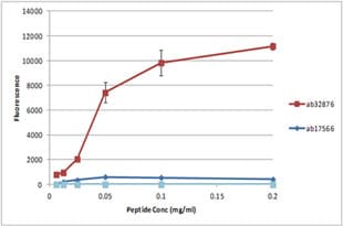
Peptide Array - Anti-Histone H3 (citrulline R2 + R8 + R17) antibody (ab5103)
All batches of ab5103 are tested in Peptide Array against peptides to different Histone H3 modifications. Six dilutions of each peptide are printed on to the Peptide Array in triplicate and results are averaged before being plotted on to a graph. Results show strong binding to Histone H3 - citrulline 2 + 8 + 17 peptide (ab32876), indicating that this antibody specifically recognises the Histone H3 - citrulline 2 + 8 + 17 modifications.
ab32876 - Histone H3 - citrulline 2 + 8 + 17
ab17566 - Histone H3 - unmodified

Western blot - Anti-Histone H3 (citrulline R2 + R8 + R17) antibody (ab5103)
All lanes : Anti-Histone H3 (citrulline R2 + R8 + R17) antibody (ab5103) at 1 µg/ml
Lane 1 : HL60 (Human Caucasian promyelocytic leukaemia) DMSO and Calcium Ionophore treated Whole Cell Lysate with with 5% BSA
Lane 2 : HL60 (Human Caucasian promyelocytic leukaemia) DMSO and Calcium Ionophore treated Whole Cell Lysate with with 5% milk
Lane 3 : HL60 (Human Caucasian promyelocytic leukaemia) DMSO and Calcium Ionophore treated Whole Cell Lysate with with 3% milk
Lysates/proteins at 10 µg per lane.
Secondary
All lanes : Goat Anti-Rabbit IgG H&L (HRP) (ab97051) at 1/10000 dilution
Developed using the ECL technique.
Performed under reducing conditions.
Predicted band size: 15 kDa
Observed band size: 17 kDawhy is the actual band size different from the predicted?
Exposure time: 30 seconds
Abcam recommends using milk as the blocking agent. Abcam welcomes customer feedback and would appreciate any comments regarding this product and the data presented above .
Blots were developled with Goat Anti-Rabbit IgG H&L (HRP) (ab97051) secondary antibody


