Anti-Paxillin (phospho Y118)抗体
参阅全部 Paxillin 一抗
兔多克隆抗体to Paxillin (phospho Y118)
Rabbit
适用于: WBmore details
与反应: Mouse, Human, African green monkey
预测可用于: Chicken![]()
Synthetic peptide corresponding to Human Paxillin (phospho Y118).
WB: COS-7, A431, HeLa whole cell lysates
The Life Science industry has been in the grips of a reproducibility crisis for a number of years. Abcam is leading the way in addressing this with our range of recombinant monoclonal antibodies and knockout edited cell lines for gold-standard validation. Please check that this product meets your needs before purchasing.
If you have any questions, special requirements or concerns, please send us an inquiry and/or contact our Support team ahead of purchase. Recommended alternatives for this product can be found below, along with publications, customer reviews and Q&As
Liquid
Shipped at 4°C. Upon delivery aliquot and store at -20°C or -80°C. Avoid repeated freeze / thaw cycles.
pH: 7.30
Preservative: 0.05% Sodium azide
Constituents: PBS, 0.1% BSA
BSA is IgG and protease free
浓度
批次浓度范围 50 µl 浓度为 0.5 - 0.8 mg/ml
Immunogen affinity purified
Purified from rabbit serum by sequential epitope-specific chromatography. The antibody has been negatively preadsorbed using i) a non-phosphopeptide corresponding to the site of phosphorylation to remove antibody that is reactive with non-phosphorylated paxillin, and ii) a generic tyrosine phosphorylated peptide to remove antibody that is reactive with phosphotyrosine, irrespective of the sequence. The final product is generated by affinity chromatography using a paxillin-derived peptide that is phosphorylated at tyrosine 31.
多克隆
IgG
Abpromise™承诺保证使用ab4833于以下的经测试应用
“应用说明”部分 下显示的仅为推荐的起始稀释度;实际最佳的稀释度/浓度应由使用者检定。
| 应用 | Ab评论 | 说明 |
|---|---|---|
| WB | 1/1000. Predicted molecular weight: 68 kDa. |
Entrez Gene: 395832 Chicken
Entrez Gene: 5829 Human
Entrez Gene: 19303 Mouse
Omim: 602505 Human
SwissProt: P49024 Chicken
SwissProt: P49023 Human
SwissProt: Q8VI36 Mouse
Unigene: 446336 Human
Unigene: 18714 Mouse
FLJ16691 antibody
FLJ23042 antibody
Paired box protein Pax 1 antibody
PAX 1 antibody
PAX1 antibody
PAXI_HUMAN antibody
Paxillin alpha antibody
Paxillin antibody
PXN antibody
PXN protein antibody

Western blot - Anti-Paxillin (phospho Y118) antibody (ab4833)
All lanes : Anti-Paxillin (phospho Y118) antibody (ab4833) at 1/500 dilution
Lane 1 : COS-7 (African green monkey kidney fibroblast-like cell line) whole cell lysate
Lane 2 : COS-7 whole cell lysate treated with 100 uM Pervanadate for 1 hour
Lane 3 : A431 (Human epidermoid carcinoma cell line) whole cell lysate
Lane 4 : A431 whole cell lysate treated with 100 uM Pervanadate for 1 hour
Lane 5 : HeLa (Human epithelial cell line from cervix adenocarcinoma) whole cell lysate
Lane 6 : HeLa whole cell lysate treated with 100 uM Pervanadate for 1 hour
Lysates/proteins at 30 µg per lane.
Secondary
All lanes : Goat anti-Rabbit IgG (H+L) Secondary Antibody, HRP conjugate at 1/5000 dilution
Predicted band size: 68 kDa
A 66 kDa band corresponding to Paxillin pTyr118 was enhanced across treated cell lines. Known quantity of protein samples were electrophoresed using Novex® NuPAGE® 12 % Bis-Tris gel, XCell SureLock™ Electrophoresis System and Novex® Sharp Pre-Stained Protein Standard. Resolved proteins were then transferred onto a nitrocellulose membrane with iBlot® 2 Dry Blotting System. The membrane was probed with the relevant primary and secondary Antibody following blocking with 5 % skimmed milk. Chemiluminescent detection was performed using Pierce™ ECL Western Blotting Substrate.
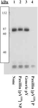
Western blot - Anti-Paxillin (phospho Y118) antibody (ab4833)
All lanes : Anti-Paxillin (phospho Y118) antibody (ab4833) at 0.75 µg/ml (3% BSA-TBST buffer)
All lanes : NMµMG (mouse mammary) cells transfected with EGFP-tagged paxillin
Secondary
All lanes : goat F(ab’)2 anti-rabbit IgG alkaline phosphatase
Predicted band size: 68 kDa
Peptide Competition: Extracts were resolved by SDS-PAGE on a 10% polyacrylamide gel and transferred to PVDF. Membranes were blocked with a 5% BSA-TBST buffer overnight at 4oC.
Prior incubation with:
1- no peptide,
2- the non-phosphopeptide corresponding to the immunogen
3- a generic phosphotyrosine containing peptide
4- the phosphopeptide immunogen
Bands were detected using the Tropix WesternStar method.
The data show that only the peptide corresponding to ab4833 blocks the antibody signal, thereby demonstrating the specificity of the antibody.


