Anti-Sumo 2 + Sumo 3抗体
参阅全部 Sumo 2 + Sumo 3 一抗
兔多克隆抗体to Sumo 2 + Sumo 3
Rabbit
Recognises 2 bands representing Sumo 2 and Sumo 3 at 15 and 18kDa in Hela Nuclear extract by Western blotting.
Replenishment batches of our polyclonal antibody, ab3742 are tested in WB. Previous batches were additionally validated in ICC/IF and IHC-P. These applications are still expected to work and are covered by our Abpromise guarantee. You may also be interested in our alternative recombinant antibody, ab81371.
适用于: ICC/IF, WB, IHC-Pmore details
与反应: Human
预测可用于: Mouse, Rat, Cow, Pig, Xenopus laevis, Zebrafish, Chinese hamster![]()
Synthetic peptide. This information is proprietary to Abcam and/or its suppliers.
WB: HeLa Nuclear cell lysate. IHC-P: Human colon adenocarcinoma tissue. ICC/IF: HepG2 cells.
The Life Science industry has been in the grips of a reproducibility crisis for a number of years. Abcam is leading the way in addressing this with our range of recombinant monoclonal antibodies and knockout edited cell lines for gold-standard validation. Please check that this product meets your needs before purchasing.
If you have any questions, special requirements or concerns, please send us an inquiry and/or contact our Support team ahead of purchase. Recommended alternatives for this product can be found below, along with publications, customer reviews and Q&As
Liquid
Shipped at 4°C. Store at +4°C short term (1-2 weeks). Upon delivery aliquot. Store at -20°C or -80°C. Avoid freeze / thaw cycle.
pH: 7.40
Preservative: 0.02% Sodium azide
Constituents: 98.98% PBS, 1% BSA
Batches of this product that have a concentration < 1mg/ml may have BSA added as a stabilising agent. If you would like information about the formulation of a specific lot, please contact our scientific support team who will be happy to help.
浓度
100 µg 浓度为 0.9 mg/ml
Immunogen affinity purified
多克隆
IgG
Abpromise™承诺保证使用ab3742于以下的经测试应用
“应用说明”部分 下显示的仅为推荐的起始稀释度;实际最佳的稀释度/浓度应由使用者检定。
| 应用 | Ab评论 | 说明 |
|---|---|---|
| ICC/IF | (3) | 1/100. |
| WB | (14) | 1/1000. Detects a band of approximately 15, 18 kDa (predicted molecular weight: 11.6 , 10.8 kDa). |
| IHC-P | (1) | 1/800. Perform heat mediated antigen retrieval before commencing with IHC staining protocol. |
Entrez Gene: 100689105 Chinese hamster
Entrez Gene: 6612 Human
Entrez Gene: 6613 Human
Entrez Gene: 170930 Mouse
Entrez Gene: 20610 Mouse
Entrez Gene: 444021 Xenopus laevis
Omim: 602231 Human
Omim: 603042 Human
SwissProt: Q6LDZ8 Chinese hamster
SwissProt: P55854 Human
SwissProt: P61956 Human
SwissProt: P61957 Mouse
SwissProt: Q9Z172 Mouse
SwissProt: Q6GPW2 Xenopus laevis
SwissProt: Q6DHL4 Zebrafish
HSMT3 antibody
MGC117191 antibody
OTTHUMP00000115275 antibody
OTTHUMP00000115276 antibody
OTTHUMP00000115277 antibody
Sentrin 2 antibody
Small ubiquitin like modifier 2 antibody
Small ubiquitin like modifier 3 antibody
Small ubiquitin related modifier 2 antibody
Small ubiquitin related modifier 3 antibody
small ubiquitin-like modifier 3 antibody
small ubiquitin-related modifier 3 antibody
SMT3 homolog 1 antibody
SMT3 homolog 2 antibody
SMT3 suppressor of mif two 3 homolog 1 antibody
SMT3 suppressor of mif two 3 homolog 2 (S. cerevisiae) antibody
SMT3 suppressor of mif two 3 homolog 2 antibody
SMT3 suppressor of mif two 3 homolog 3 (S. cerevisiae) antibody
SMT3 suppressor of mif two 3 homolog 3 antibody
SMT3A antibody
SMT3B antibody
SMT3H1 antibody
SMT3H2 antibody
Sumo2 antibody
Sumo3 antibody
Ubiquitin like protein SMT3A antibody
Ubiquitin like protein SMT3B antibody
Ubiquitin-like protein SMT3B antibody

Immunocytochemistry/ Immunofluorescence - Anti-Sumo 2 + Sumo 3 antibody (ab3742)
ab3742 staining Sumo 2 + Sumo 3 in HepG2 cells. The cells were fixed with 4% paraformaldehyde (10 min), permeabilized with 0.1% PBS-Triton X-100 for 5 minutes and then blocked with 1% BSA/10% normal goat serum/0.3M glycine in 0.1%PBS-Tween for 1h. The cells were then incubated overnight at 4°C with ab3742 at 1µg/ml and ab7291, Mouse monoclonal [DM1A] to alpha Tubulin - Loading Control. Cells were then incubated with ab150081, Goat polyclonal Secondary Antibody to Rabbit IgG - H&L (Alexa Fluor® 488), pre-adsorbed at 1/1000 dilution (shown in green) and ab150120, Goat polyclonal Secondary Antibody to Mouse IgG - H&L (Alexa Fluor® 594), pre-adsorbed at 1/1000 dilution (shown in pseudocolour red). Nuclear DNA was labelled with DAPI (shown in blue).
Image was acquired with a high-content analyser (Operetta CLS, Perkin Elmer) and a maximum intensity projection of confocal sections is shown.

Immunocytochemistry/ Immunofluorescence - Anti-Sumo 2 + Sumo 3 antibody (ab3742)Image from Cuchet-Lourenço D et al., PLoS Pathog. 2011;7(7):e1002123. Fig 9(B).; doi: 10.1371/journal.ppat.1002123. Reproduced under the Creative Commons license http://creativecommons.org/licenses/by/4.0/
Left-hand images show uninfected cells and the co-localization of SUMO-2/3 with PML (red) in control (upper rows of each block of 4 images) and PML depleted (low rows of each block of 4 images) HepaRG cells. Right-hand images show typical examples of recruitment of the indicated proteins to sites associated with HSV-1 genomes (ICP4; red) in cells at the edges of ICP0 null mutant (ΔICP0) plaques in control and PML depleted HepaRG cells. Scale bars indicate 5 µm.
Cells on glass coverslips were fixed with 1.5% formaldehyde in PBS containing 2% sucrose then treated with 0.5% Nonidet P40 substitute in PBS/10% sucrose. SUMO-2/3 was detected with ab3742. An Alexa-conjugated anti-rabbit IgG was used as the secondary antibody.

Western blot - Anti-Sumo 2 + Sumo 3 antibody (ab3742)
Lanes 1 & 3: Sumo 2+3 antibody (ab3742) at 1/500 dilution
Lanes 2 & 4: Sumo 2+3 antibody (ab3742) at 1/1000 dilution
Lanes 1-4: HeLa nuclear extract at 20 ug
Lane 1: as above
Lane 2: as above
Lane 3: Sumo 2+3 peptide (ab13760) at 1 ug/ml
Lane 4: Sumo 2+3 peptide (ab13760) at 1 ug/ml
Secondary
Goat polyclonal to Rabbit IgG H&L (HRP) (ab6721) at 1/2000 dilution developed using the ECL technique
Performed under reducing conditions.
Exposure time: 1 minute
Predicted band sizes : 11.6 & 10.

Immunohistochemistry (Formalin/PFA-fixed paraffin-embedded sections) - Anti-Sumo 2 + Sumo 3 antibody (ab3742)
IHC image of Sumo 2+3 staining in human colon adenocarcinoma formalin fixed paraffin embedded tissue section, performed on a Leica BondTM system using the standard protocol F. The section was pre-treated using heat mediated antigen retrieval with sodium citrate buffer (pH6, epitope retrieval solution 1) for 20 mins. The section was then incubated with ab3742, 1µg/ml, for 15 mins at room temperature and detected using an HRP conjugated compact polymer system. DAB was used as the chromogen. The section was then counterstained with haematoxylin and mounted with DPX.
For other IHC staining systems (automated and non-automated) customers should optimize variable parameters such as antigen retrieval conditions, primary antibody concentration and antibody incubation times.

Immunohistochemistry (Formalin/PFA-fixed paraffin-embedded sections) - Anti-Sumo 2 + Sumo 3 antibody (ab3742)Image courtesy of Human Protein Atlas
Image courtesy of Human Protein Atlas
ab3742 staining Sumo 2 + 3 in Human skin. The paraffin embedded human skin tissue was incubated with ab3742 (1/800 dilution) for 30 mins at room temperature. Antigen retrieval was performed by heat induction in citrate buffer pH 6. Ab3742 was tested in a tissue microarray (TMA) containing a wide range of normal and cancer tissues as well as a cell microarray consisting of a range of commonly used, well characterised human cell lines.
Further results for this antibody can be found at www.proteinatlas.org

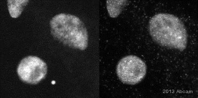
Immunocytochemistry/ Immunofluorescence - Anti-Sumo 2 + Sumo 3 antibody (ab3742)This image is courtesy of Luke Hughes-Davies and Rhiannon Jade, Gurdon Institute, Cambridge, UK
Immunofluorescent imaging of human cells (U2OS) with ab3742 confirms the specificity of this antibody. Antibody signal is localised exclusively to the nucleus, with diffuse background staining of the nucleoplasm. Intense foci of staining are also evident, corresponding to SUMO-2/3 accumulation in nuclear subdomains such as the PML body. This image is in exact agreement with several published reports (see for example Saitoh H et al.).
IF was performed with a standard paraformaldehyde technique (fixed in PBS buffered PFH 4% for 5 minutes, permeabilised with 0.5% triton-PBS for 5 minutes, blocked with 5% milk / 0.2% tween for one hour. Primary antibody used at 1/100 in 5% milk / 0.2% TWEEN for one hour, secondary antibody for 30 minutes. All blocking and incubation steps carried out at 37 degrees. Nuclei are stained with Hoechst stain.
Immunocytochemistry/ Immunofluorescence - Anti-Sumo 2 + Sumo 3 antibody (ab3742)This image is courtesy of an Abreview submitted by Andrew Lane
ab3742 staining Sumo 2+3 in Xenopus laevis oocyte cell by ICC/IF (Immunocytochemistry/immunofluorescence). Cells were fixed with formaldehyde, permeabilized with NP-40 0.1% in PBS and blocked with 3% BSA for 60 minutes at 23°C. Samples were incubated with primary antibody (1/250 in PBS + 3% BSA) for 1 hour at 23°C. An undiluted Alexa Fluor®488-conjugated Goat anti-rabbit polyclonal was used as the secondary antibody.


