Anti-Endostatin/COL18A1抗体
参阅全部 Endostatin/COL18A1 一抗
兔多克隆抗体to Endostatin/COL18A1
Rabbit
Detects recombinant human Endostatin/COL18A1.
适用于: ICC/IF, IHC-Pmore details
与反应: Mouse, Cat, Human, Non human primates
This product was produced with the following immunogens:
Synthetic peptide corresponding to Mouse Endostatin/COL18A1.
RRLMESYCETWRTE
Database link: P39061
Synthetic peptide corresponding to Human Endostatin/COL18A1. RRLTESYCETWRTE
Database link: P39060
ab3453 has been recombinant only tested in Western blot.
The Life Science industry has been in the grips of a reproducibility crisis for a number of years. Abcam is leading the way in addressing this with our range of recombinant monoclonal antibodies and knockout edited cell lines for gold-standard validation. Please check that this product meets your needs before purchasing.
If you have any questions, special requirements or concerns, please send us an inquiry and/or contact our Support team ahead of purchase. Recommended alternatives for this product can be found below, along with publications, customer reviews and Q&As
Liquid
Shipped at 4°C. Store at +4°C short term (1-2 weeks). Upon delivery aliquot. Store at -20°C or -80°C. Avoid freeze / thaw cycle.
Preservative: 0.05% Sodium azide
Constituents: 0.1% BSA, 99% PBS
浓度
100 µg 浓度为 1 mg/ml
Immunogen affinity purified
多克隆
IgG
Abpromise™承诺保证使用ab3453于以下的经测试应用
“应用说明”部分 下显示的仅为推荐的起始稀释度;实际最佳的稀释度/浓度应由使用者检定。
| 应用 | Ab评论 | 说明 |
|---|---|---|
| ICC/IF | 1/200 - 1/1000. | |
| IHC-P | 1/100 - 1/500. |
Entrez Gene: 1306 Human
Entrez Gene: 80781 Human
Entrez Gene: 12819 Mouse
Entrez Gene: 12822 Mouse
Omim: 120328 Human
SwissProt: P39059 Human
SwissProt: P39060 Human
SwissProt: O35206 Mouse
SwissProt: P39061 Mouse
Unigene: 409034 Human
Unigene: 517356 Human
Unigene: 233547 Mouse
Unigene: 4352 Mouse
Alpha 1 collagen type 18 (XVIII)(COL18A1) antibody
Alpha 1 type XVIII collagen antibody
Antiangiogenic agent antibody
COIA1_HUMAN antibody
COL15A1 antibody
Col18a1 antibody
Collagen alpha 1(XV) chain antibody
Collagen alpha 1(XVIII) chain antibody
Collagen alpha-1(XV) chain antibody
Collagen type XV proteoglycan antibody
Collagen type XVIII alpha 1 antibody
Collagen XV, alpha 1 polypeptide antibody
Collagen, type XV, alpha 1 antibody
Endostatin antibody
Endostatin XV antibody
FLJ27325 antibody
FLJ34914 antibody
FLJ38566 antibody
KNO antibody
KNO1 antibody
KS antibody
MGC74745 antibody
Multi functional protein MFP antibody
OTTHUMP00000021782 antibody
OTTHUMP00000115472 antibody
OTTHUMP00000115473 antibody

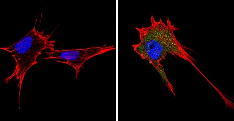
Immunocytochemistry/ Immunofluorescence - Anti-Endostatin/COL18A1 antibody (ab3453)
ab3453 labelling Endostatin/COL18A1 (green) in 293T cells (right) compared with a negative control (left) by Immunocytochemistry/Immunofluorscence. Formalin-fixed cells were permeabilized with 0.1% Triton X-100 in TBS for 5-10 minutes, blocked with 3% BSA-PBS for 30 minutes at room temperature. Cells were incubated with the primary antibody (1:200 in 3% BSA-PBS) overnight at 4 ºC. A DyLight 488-conjugated Goat anti-rabbit IgG was used as the secondary antibody. Red (phalloidin) - F-actin, Blue (DAPI) - nuclei (blue). Images were taken at a magnification of 60x.

Immunocytochemistry/ Immunofluorescence - Anti-Endostatin/COL18A1 antibody (ab3453)
ab3453 labelling Endostatin/COL18A1 (green) in A431 cells (right) compared with a negative control (left) by Immunocytochemistry/Immunofluorscence. Formalin-fixed cells were permeabilized with 0.1% Triton X-100 in TBS for 5-10 minutes, blocked with 3% BSA-PBS for 30 minutes at room temperature. Cells were incubated with the primary antibody (1:200 in 3% BSA-PBS) overnight at 4 ºC. A DyLight 488-conjugated Goat anti-rabbit IgG was used as the secondary antibody. Red (phalloidin) - F-actin, Blue (DAPI) - nuclei (blue). Images were taken at a magnification of 60x.
Immunocytochemistry/ Immunofluorescence - Anti-Endostatin/COL18A1 antibody (ab3453)
ab3453 labelling Endostatin/COL18A1 (green) in NIH-3T3 cells (right) compared with a negative control (left) by Immunocytochemistry/Immunofluorscence. Formalin-fixed cells were permeabilized with 0.1% Triton X-100 in TBS for 5-10 minutes, blocked with 3% BSA-PBS for 30 minutes at room temperature. Cells were incubated with the primary antibody (1:200 in 3% BSA-PBS) overnight at 4 ºC. A DyLight 488-conjugated Goat anti-rabbit IgG was used as the secondary antibody. Red (phalloidin) - F-actin, Blue (DAPI) - nuclei (blue). Images were taken at a magnification of 60x.

Immunohistochemistry (Formalin/PFA-fixed paraffin-embedded sections) - Anti-Endostatin/COL18A1 antibody (ab3453)
ab3453 labelling Endostatin/COL18A1 in the secretion of Human kidney tissue (right) compared with a negative control (left) by Immunohistochemistry (formalin/PFA-fixed paraffin embedded sections). To expose target proteins, antigen retrieval method was performed using 10mM sodium citrate (pH 6.0), microwaved for 8-15 min. Following antigen retrieval, tissues were blocked in 3% H2O2-methanol for 15 min at room temperature. Tissue sections were incuabted with the primary antibody (1:100 in 3% BSA-PBS) overnight at 4°C. A HRP-conjugated anti-rabbit IgG was used as the secondary antibody, followed by colorimetric detection using a DAB kit. Tissues were counterstained with hematoxylin and dehydrated with ethanol and xylene to prep for mounting.

Immunohistochemistry (Formalin/PFA-fixed paraffin-embedded sections) - Anti-Endostatin/COL18A1 antibody (ab3453)
ab3453 labelling Endostatin/COL18A1 in the secretion of Human colon tissue (right) compared with a negative control (left) by Immunohistochemistry (formalin/PFA-fixed paraffin embedded sections). To expose target proteins, antigen retrieval method was performed using 10mM sodium citrate (pH 6.0), microwaved for 8-15 min. Following antigen retrieval, tissues were blocked in 3% H2O2-methanol for 15 min at room temperature. Tissue sections were incuabted with the primary antibody (1:200 in 3% BSA-PBS) overnight at 4°C. A HRP-conjugated anti-rabbit IgG was used as the secondary antibody, followed by colorimetric detection using a DAB kit. Tissues were counterstained with hematoxylin and dehydrated with ethanol and xylene to prep for mounting.

Immunohistochemistry (Formalin/PFA-fixed paraffin-embedded sections) - Anti-Endostatin/COL18A1 antibody (ab3453)
ab3453 labelling Endostatin/COL18A1 in the secretion of Mouse kidney tissue (right) compared with a negative control (left) by Immunohistochemistry (formalin/PFA-fixed paraffin embedded sections). To expose target proteins, antigen retrieval method was performed using 10mM sodium citrate (pH 6.0), microwaved for 8-15 min. Following antigen retrieval, tissues were blocked in 3% H2O2-methanol for 15 min at room temperature. Tissue sections were incuabted with the primary antibody (1:100 in 3% BSA-PBS) overnight at 4°C. A HRP-conjugated anti-rabbit IgG was used as the secondary antibody, followed by colorimetric detection using a DAB kit. Tissues were counterstained with hematoxylin and dehydrated with ethanol and xylene to prep for mounting.


