Anti-KAT13D / CLOCK抗体
参阅全部 KAT13D / CLOCK 一抗
兔多克隆抗体to KAT13D / CLOCK
Rabbit
适用于: IHC-P, ICC/IF, IHC-Fr, WB, ChIPmore details
与反应: Mouse, Human
Synthetic peptide corresponding to Mouse KAT13D/ CLOCK aa 1-100.
WB: human HeLa, mouse NIH-3T3 and skeletal muscle tissue; IHC: human skeletal muscle, colon tissue, mouse colon tissue; ICC: human U251 cells
The Life Science industry has been in the grips of a reproducibility crisis for a number of years. Abcam is leading the way in addressing this with our range of recombinant monoclonal antibodies and knockout edited cell lines for gold-standard validation. Please check that this product meets your needs before purchasing.
If you have any questions, special requirements or concerns, please send us an inquiry and/or contact our Support team ahead of purchase. Recommended alternatives for this product can be found below, along with publications, customer reviews and Q&As
Liquid
Shipped at 4°C. Store at +4°C short term (1-2 weeks). Upon delivery aliquot. Store at -20°C or -80°C. Avoid freeze / thaw cycle.
Preservative: 0.05% Sodium azide
Constituents: 0.1% BSA, 99% PBS
Immunogen affinity purified
多克隆
IgG
Abpromise™承诺保证使用ab3517于以下的经测试应用
“应用说明”部分 下显示的仅为推荐的起始稀释度;实际最佳的稀释度/浓度应由使用者检定。
| 应用 | Ab评论 | 说明 |
|---|---|---|
| IHC-P | (1) | 1/100 - 1/1000. Immunohistochemical staining of CLOCK in hamster brain results in the staining of the superchiasmatic nucleus. |
| ICC/IF | (1) | 1/10 - 1/200. |
| EMSA | Use at an assay dependent concentration. | |
| IHC-Fr | Use at an assay dependent concentration. | |
| Gel Shift Assay | Use at an assay dependent concentration. | |
| WB | (3) | 1/200 - 1/2000. Detects a band of approximately 100 kDa (predicted molecular weight: 95 kDa). |
| ChIP | Use at an assay dependent concentration. PubMed: 20956306 |
Entrez Gene: 9575 Human
Entrez Gene: 12753 Mouse
Omim: 601851 Human
SwissProt: O15516 Human
SwissProt: O08785 Mouse
Unigene: 436975 Human
Unigene: 3552 Mouse
Unigene: 392894 Mouse
Circadian Locomotor Output Cycles Kaput antibody
Circadium Locomotor Output Cycles Kaput antibody
Class E basic helix-loop-helix protein 8 antibody
CLOCK antibody
Clock circadian regulator antibody
Clock homolog antibody
Clock protein antibody
CLOCK_HUMAN antibody
hCLOCK antibody
KIAA0334 antibody
bHLHe8 antibody
Circadian locomoter output cycles kaput protein antibody
Circadian locomoter output cycles protein kaput antibody

Western blot - Anti-KAT13D / CLOCK antibody (ab3517)
All lanes : Anti-KAT13D / CLOCK antibody (ab3517) at 1/2000 dilution
Lane 1 : Mouse skeletal muscle tissue
Lane 2 : Mouse liver tissue
Lysates/proteins at 30 µg per lane.
Secondary
Lane 1 : Goat anti-Rabbit IgG (H+L) Superclonal™ Recombinant Secondary Antibody, HRP at 1/4000 dilution
Lane 2 : oat anti-Rabbit IgG (H+L) Superclonal™ Recombinant Secondary Antibody, HRP at 1/4000 dilution
Developed using the ECL technique.
Predicted band size: 95 kDa
Electrophoresis performed on a 4-12% BisTris gel and proteins transferred onto a nitrocellulose membrane.

Immunocytochemistry/ Immunofluorescence - Anti-KAT13D / CLOCK antibody (ab3517)
Immunocytochemistry/immunofluorescence analysis of U251 cells labeling KAT13D/CLOCK (green) with ab3517 at 1/100. Cells were fixed with formalin and permeabilized with 0.1% Triton X-100 in TBS for 5-10 minutes and blcoked with £% BSA in PBS for 30 minutes at room temperature. Cells were incubated with the primary antibody overnight at 4°C. A DyLight-conjugated secondary antibody was used. F-actin (red) was stained with phalloidin and nuclei (blue) were stained with Hoechst or DAPI. 60X magnification. Left - negative control.

Immunohistochemistry (Formalin/PFA-fixed paraffin-embedded sections) - Anti-KAT13D / CLOCK antibody (ab3517)
ab3517 labelling KAT13D in the nucleus and cytoplasm of Human skeletal muscle tissue (right) compared with a negative control (left) by Immunohistochemistry (formalin/PFA-fixed paraffin-embedded sections). To expose target proteins, antigen retrieval method was performed using 10mM sodium citrate (pH 6.0) microwaved for 8-15 min. Following antigen retrieval, tissues were blocked in 3% H2O2-methanol for 15 min at room temperature. Thissue sections were incubated with the primary antibody (1:200 in 3% BSA-PBS) overnight at 4°C. A HRP-conjugated anti-rabbit IgG was used as the secondary antibody, followed by colorimetric detection using a DAB kit. Tissues were counterstained with hematoxylin and dehydrated with ethanol and xylene to prep for mounting.
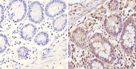
Immunohistochemistry (Formalin/PFA-fixed paraffin-embedded sections) - Anti-KAT13D / CLOCK antibody (ab3517)
ab3517 labelling KAT13D in the nucleus and cytoplasm of Mouse colon tissue (right) compared with a negative control (left) by Immunohistochemistry (formalin/PFA-fixed paraffin-embedded sections). To expose target proteins, antigen retrieval method was performed using 10mM sodium citrate (pH 6.0) microwaved for 8-15 min. Following antigen retrieval, tissues were blocked in 3% H2O2-methanol for 15 min at room temperature. Thissue sections were incubated with the primary antibody (1:200 in 3% BSA-PBS) overnight at 4°C. A HRP-conjugated anti-rabbit IgG was used as the secondary antibody, followed by colorimetric detection using a DAB kit. Tissues were counterstained with hematoxylin and dehydrated with ethanol and xylene to prep for mounting.
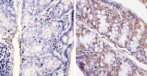
Immunohistochemistry (Formalin/PFA-fixed paraffin-embedded sections) - Anti-KAT13D / CLOCK antibody (ab3517)
ab3517 labelling KAT13D in the nucleus and cytoplasm of Human colon tissue (right) compared with a negative control (left) by Immunohistochemistry (formalin/PFA-fixed paraffin-embedded sections). To expose target proteins, antigen retrieval method was performed using 10mM sodium citrate (pH 6.0) microwaved for 8-15 min. Following antigen retrieval, tissues were blocked in 3% H2O2-methanol for 15 min at room temperature. Thissue sections were incubated with the primary antibody (1:200 in 3% BSA-PBS) overnight at 4°C. A HRP-conjugated anti-rabbit IgG was used as the secondary antibody, followed by colorimetric detection using a DAB kit. Tissues were counterstained with hematoxylin and dehydrated with ethanol and xylene to prep for mounting.

Immunohistochemistry (Formalin/PFA-fixed paraffin-embedded sections) - Anti-KAT13D / CLOCK antibody (ab3517)This image is courtesy of an anonymous Abreview
ab3517 staining KAT13D/CLOCK in Mouse skeletal muscle tissue sections by IHC-P (Paraformaldehyde-fixed, paraffin-embedded tissue sections). Tissue was fixed with paraformaldehyde and blocked with 10% serum for 1 hour at 20°C; antigen retrieval was by heat mediation in citrate buffer pH6. Samples were incubated with primary antibody (1/400 in PBS) for 12 hours at 4°C. Undiluted ab64256 was used as the secondary antibody.

Western blot - Anti-KAT13D / CLOCK antibody (ab3517)
All lanes : Anti-KAT13D / CLOCK antibody (ab3517) at 1/4000 dilution
Lane 1 : HeLa cell lysate
Lane 2 : NIH-3T3 cell lysate
Lysates/proteins at 25 µg per lane.
Predicted band size: 95 kDa
Observed band size: 100 kDawhy is the actual band size different from the predicted?


