Anti-CD45抗体
参阅全部 CD45 一抗
兔多克隆抗体to CD45
Rabbit
适用于: WB, IHC-P, Flow Cyt (Intra)more details
与反应: Mouse, Rat, Human
预测可用于: Pig, Rhesus monkey![]()
Synthetic peptide. This information is proprietary to Abcam and/or its suppliers.
The Life Science industry has been in the grips of a reproducibility crisis for a number of years. Abcam is leading the way in addressing this with our range of recombinant monoclonal antibodies and knockout edited cell lines for gold-standard validation. Please check that this product meets your needs before purchasing.
If you have any questions, special requirements or concerns, please send us an inquiry and/or contact our Support team ahead of purchase. Recommended alternatives for this product can be found below, along with publications, customer reviews and Q&As
Liquid
Shipped at 4°C. Upon delivery aliquot and store at -20°C or -80°C. Avoid repeated freeze / thaw cycles.
pH: 7.40
Preservative: 0.02% Sodium azide
Constituents: 98.98% PBS, 1% BSA
浓度
批次浓度范围 100 µg 浓度为 0.5 - 1 mg/ml
Immunogen affinity purified
多克隆
IgG
Abpromise™承诺保证使用ab10558于以下的经测试应用
“应用说明”部分 下显示的仅为推荐的起始稀释度;实际最佳的稀释度/浓度应由使用者检定。
| 应用 | Ab评论 | 说明 |
|---|---|---|
| WB | (9) | 1/500. Detects a band of approximately 190 kDa (predicted molecular weight: 147 kDa). |
| IHC-P | (37) | Use a concentration of 0.5 - 5 µg/ml. Perform heat mediated antigen retrieval before commencing with IHC staining protocol. |
| Flow Cyt (Intra) | Use 1µg for 106 cells. ab171870 - Rabbit polyclonal IgG, is suitable for use as an isotype control with this antibody. |
Entrez Gene: 5788 Human
Entrez Gene: 19264 Mouse
Omim: 151460 Human
SwissProt: P08575 Human
SwissProt: P06800 Mouse
Unigene: 654514 Human
Unigene: 391573 Mouse
Unigene: 90166 Rat
B220 antibody
CD 45 antibody
CD45 antibody
CD45 antigen antibody
CD45R antibody
GP180 antibody
L-CA antibody
LCA antibody
Leukocyte common antigen antibody
loc antibody
Ly-5 antibody
LY5 antibody
Ly5, homolog of antibody
Lyt-4 antibody
OTTHUMP00000033813 antibody
OTTHUMP00000033816 antibody
OTTHUMP00000033817 antibody
OTTHUMP00000038574 antibody
Protein tyrosine phosphatase receptor type c polypeptide antibody
Protein tyrosine phosphatase, receptor type C antibody
protein tyrosine phosphatase, receptor type, C antibody
Protein tyrosine phosphatase, receptor type, c polypeptide antibody
Ptprc antibody
PTPRC_HUMAN antibody
Receptor-type tyrosine-protein phosphatase C antibody
T200 antibody
T200 glycoprotein antibody
T200 leukocyte common antigen antibody

Immunohistochemistry (Formalin/PFA-fixed paraffin-embedded sections) - Anti-CD45 antibody (ab10558)
Immunohistochemical analysis of formalin fixed paraffin embedded human tonsil labelling CD45 with ab10558 at a concentration of 0.1µg/ml. The immunostaining was performed on a Ventana DISCOVERY ULTRA (Roche Tissue Diagnostics) instrument with a OptiView DAB IHC Detection Kit. Heat mediated antigen retrieval was performed with DISCOVERY cell conditioning solution (CC1) 100°C, pH8.5 for 32mins.
ab10558 anti-CD45 antibody was incubated for 16mins at 37°C. Sections were counterstained with Hematoxylin II. Image inset shows absence of staining in secondary antibody only control.
Customers are encouraged to optimise antigen retrieval conditions, antibody concentration, incubation times and temperature for best results in their own IHC assay workflow (automated and manual)

Immunohistochemistry (Formalin/PFA-fixed paraffin-embedded sections) - Anti-CD45 antibody (ab10558)
Immunohistochemical analysis of formalin fixed paraffin embedded human tonsil labelling CD45 with ab10558 at a concentration of 0.5µg/ml. The immunostaining was performed on a Leica Biosystems BOND® RX instrument with a Bond™ Polymer Refine Detection kit. Heat mediated antigen retrieval was performed with Tris-EDTA buffer (pH 9.0, Epitope Retrieval Solution 2) for 20mins.
ab10558 anti-CD45 antibody was incubated for 15mins at room temperature. Sections were counterstained with Hematoxylin. Image inset shows absence of staining in secondary antibody only control.
Customers are encouraged to optimise antigen retrieval conditions, antibody concentration, incubation times and temperature for best results in their own IHC assay workflow (automated and manual)

Immunohistochemistry (Formalin/PFA-fixed paraffin-embedded sections) - Anti-CD45 antibody (ab10558)
ab10558 (1:40) staining CD45 in paraffin-embedded human tonsil (left panel) using an automated system (Ventana Discovery). Right-hand panel shows negative control (no primary antibody).
Using this protocol there is strong membrane staining of B cells in the germinal centres and mantle zone of the follicles and scattered cells of the interfollicular areas (paracortical T and B cells). There is a mild to moderate degree of cytoplasmic staining associated with the membrane staining in these specific cells.
Sections were rehydrated and antigen retrieved in CC1 Cell Conditioning Buffer using Ventana Extended Retrieval programme. Slides were blocked in 3% H2O2 /4 min/ 37°C and incubated with ab10558 (1:40 dilution / 1 hour/ 37°C). Sections then blocked (4mins/ 37°C) and incubated with Dako swine anti-rabbit antibody (1:50, 28 min/ 37°C). Staining was amplified and detected by incubation with Ventana Streptavidin ABC (HRP-DAB) system (16 min/ 37°C) before being counterstained with hematoxylin.

Immunohistochemistry (Formalin/PFA-fixed paraffin-embedded sections) - Anti-CD45 antibody (ab10558)Lorenzi T et al., PLos One 7:e35232 (2012), Fig 4, doi: 10.1371/journal.pone.0035232 Reproduced under the Creative Commons license http://creativecommons.org/licenses/by/4.0/
ab10558 staining CD45 in Human tonsil tissue sections by Immunohistochemistry (IHC-P - paraformaldehyde-fixed, paraffin-embedded sections). Negative control is shown in panel. Blocking was with horse serum (1/75) for 1 hour at room temperature. Samples were incubated with primary antibody (1/10) overnight at 4°C. A Biotin-conjugated Horse anti-mouse polyclonal (1/200) was used as the secondary antibody.

Flow Cytometry (Intracellular) - Anti-CD45 antibody (ab10558)
Overlay histogram showing Jurkat cells stained with ab10558 (red line). The cells were fixed with 80% methanol (5 min) and incubated in 1x PBS / 10% normal goat serum / 0.3M glycine to block non-specific protein-protein interactions. The cells were then incubated with the antibody (ab10558, 1µg/1x106 cells) for 30 min at 22ºC. The secondary antibody used was DyLight® 488 goat anti-rabbit IgG (H+L) (ab96899) at 1/1000 dilution for 30 min at 22ºC. Isotype control antibody (black line) was rabbit IgG (1µg/1x106 cells) used under the same conditions. Acquisition of >5,000 events was performed.
Please note that Abcam do not have any data for use of this antibody on non-fixed cells. We welcome any customer feedback.

Western blot - Anti-CD45 antibody (ab10558)
All lanes : Anti-CD45 antibody (ab10558) at 1/500 dilution
Lane 1 : Jurkat Whole Cell Lysate
Lane 2 : Jurkat Whole Cell Lysate with Human CD45 peptide (ab17553)
Lysates/proteins at 20 µg per lane.
Secondary
All lanes : Goat Anti-Rabbit IgG H&L (HRP) (ab6721) at 1/5000 dilution
Predicted band size: 147 kDa
Exposure time: 3 minutes

Immunohistochemistry (Formalin/PFA-fixed paraffin-embedded sections) - Anti-CD45 antibody (ab10558)
IHC image of CD45 antibody staining in a section of formalin-fixed paraffin-embedded normal human spleen* performed on a Leica BONDTM system using the standard protocol. The section was pre-treated using heat mediated antigen retrieval with sodium citrate buffer (pH6, epitope retrieval solution 1) for 20mins. The section was then incubated with ab10558, 1ug/ml, for 15 mins at room temperature and detected using an HRP conjugated compact polymer system. DAB was used as the chromogen. The section was then counterstained with haematoxylin and mounted with DPX.
For other IHC staining systems (automated and non-automated) customers should optimize variable parameters such as antigen retrieval conditions, primary antibody concentration and antibody incubation times.
*Tissue obtained from the Human Research Tissue Bank, supported by the NIHR Cambridge Biomedical Research Centre
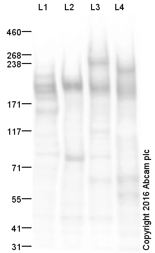
Western blot - Anti-CD45 antibody (ab10558)
All lanes : Anti-CD45 antibody (ab10558) at 1 µg/ml
Lane 1 : Jurkat (Human) Whole Cell Lysate
Lane 2 : RAW 264.7 (Mouse leukaemic monocyte macrophage cell line) Whole Cell Lysate
Lane 3 : Spleen (Mouse) Tissue Lysate
Lane 4 : Spleen (Rat) Tissue Lysate
Lysates/proteins at 20 µg per lane.
Secondary
All lanes : Goat Anti-Rabbit IgG H&L (HRP) at 1/50000 dilution
Developed using the ECL technique.
Performed under reducing conditions.
Predicted band size: 147 kDa
Observed band size: 190 kDawhy is the actual band size different from the predicted?
Additional bands at: 230 kDa. We are unsure as to the identity of these extra bands.
Exposure time: 1 minute
This blot was produced using a 3-8% Tris Acetate gel under the TA buffer system. The gel was run at 150V for 60 minutes before being transferred onto a Nitrocellulose membrane at 30V for 70 minutes. The membrane was then blocked for an hour using 2% Bovine Serum Albumin before being incubated with ab10558 overnight at 4°C. Antibody binding was detected using an anti-rabbit antibody conjugated to HRP, and visualised using ECL development solution ab133406.
CD45 contains a number of potential glycosylation sites (SwissProt) which may explain its migration at a higher molecular weight than predicted.

Flow Cytometry (Intracellular) - Anti-CD45 antibody (ab10558)This image is courtesy of an abreview submitted by Kirk Mcmanu.
Asynchronous KM-H2 cells were pelleted and labeled by indirect immunofluorescence. Cells were stained with ab10558 (1/200) for 30min at 4'C, washed and then stained with goat anti-rabbit alexafluor 488 (1/200). Forward/Side scatter were used to eliminate cellular debris. The accompanying marker was applied such that only 2% of the IgG control was positive Based on the accompanying image, approximately 8.4% of cells exhibited positive staining for anti-CD45. Since KM-H2 are known to have low levels of CD45 transcripts they are expected to have low levels of CD45, which is reflected in the ~8%. This image is from an Abreview.

Immunohistochemistry (Formalin/PFA-fixed paraffin-embedded sections) - Anti-CD45 antibody (ab10558)This image is courtesy of an anonymous abreview.
Immunohistochemical analysis of formaldehyde fixed human cephalic sections. Primary antibody ab10558 to CD45 incubated at a concentration of 1/100 for 4°C for 18 hours. Secondary antibody used was a goat anti-rabbit congugated to biotin at a 1/200 dilution. Blocking was done with serum at a 10% concentration for 1 hour at 25°C.


