| 存储条件 |
|---|
| Product Name | RAB27A Rabbit mAb |
|---|---|
| Antibody Type | Primary Antibodies |
| Immunogen | Recombinant fusion protein containing a sequence corresponding to amino acids 1-221 of human RAB27A(NP_004571.2). |
| Clonality | monoclonal |
|---|---|
| Isotype | IgG |
| Host Species | Rabbit |
| Tested Applications | ICC/IFIHCWB |
| WB:1:1500-1:6000 IHC:1:200-1:2000 ICC/IF: 1:200-1:800 | |
| Species Reactivity | HumanMouseRat |
| Concentration | 1mg/ml |
| Purification | Affinity purified |
| Gene Symbol | RAB27A |
|---|---|
| Gene Synonyms | GS2 RAM RAB27 HsT18676 |
| Gene Full Name | RAB27A, member RAS oncogene family |
| Gene Summary | The protein encoded by this gene belongs to the small GTPase superfamily, Rab family. The protein is membrane-bound and may be involved in protein transport and small GTPase mediated signal transduction. Mutations in this gene are associated with Griscelli syndrome type 2. Alternative splicing occurs at this locus and four transcript variants encoding the same protein have been identified. [provided by RefSeq, Jul 2008] |
| Alternative Names | GS2 RAM RAB27 HsT18676 RAB27A |
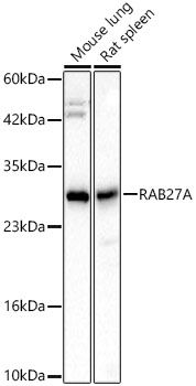
| Molecular Weight(MW) | 25kDa(Observed MW 27kDa) |
| Cellular Localization | Late endosome, Lipid-anchor, Lysosome, Melanosome, Membrane. |
WB
Western blot analysis of various lysates, using RAB27A Rabbit mAb at 1:3000 dilution. Secondary antibody: HRP-conjugated Goat anti-Rabbit IgG (H+L) at 1:10000 dilution. Lysates/proteins: 25μg per lane. Blocking buffer: 3% nonfat dry milk in TBST. Detection: ECL Basic Kit. Exposure time: 45s.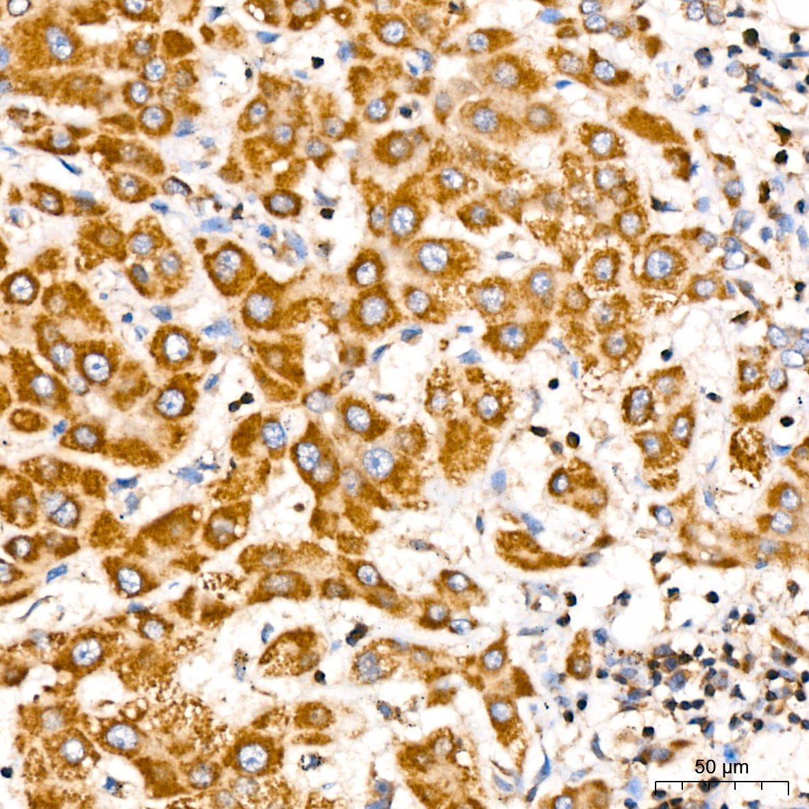
IHC
Immunohistochemistry analysis of paraffin embedded Human liver using RAB27A Rabbit mAb at dilution of 1:300 (40x lens). High pressure antigen retrieval performed with 0.01M Citrate Bufferr (pH 6.0) prior to IHC staining.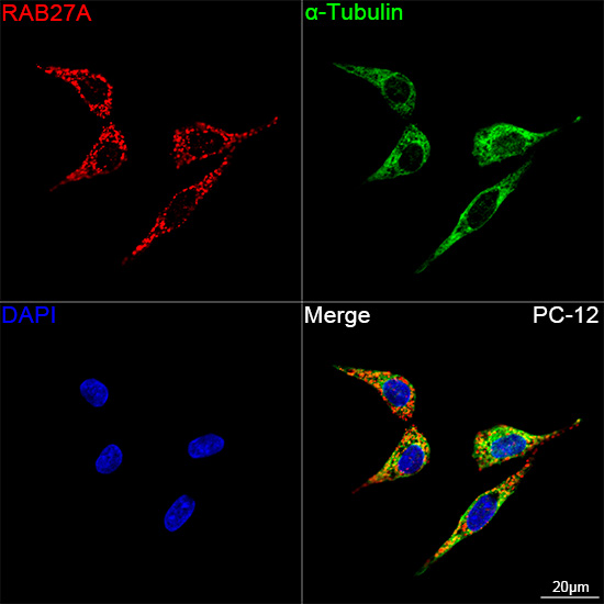
ICC/IF
Confocal imaging of PC-12 cells using RAB27A Rabbit mAb (dilution 1:200) followed by a further incubation with Cy3 Goat Anti-Rabbit IgG (H+L) (dilution 1:500) (Red). The cells were counterstained with α-Tubulin Mouse mAb (dilution 1:400) followed by incubation with 488-conjugated Goat Anti-Mouse IgG (H+L) Ab (dilution 1:500) (Green). DAPI was used for nuclear staining (Blue). Objective: 100x.| Application Notes | WB:1:1500-1:6000 IHC:1:200-1:2000 ICC/IF: 1:200-1:800 |
|---|
| Form | Liquid |
|---|---|
| Storage Instructions | Store at -20℃. Avoid freeze / thaw cycles. |
| Storage Buffer | Buffer: PBS with 0.05% proclin300, 0.05% BSA, 50% glycerol, pH7.3. |
抱歉,暂无相关文献







