| 存储条件 |
|---|
| Product Name | UBE3A Rabbit pAb |
|---|---|
| Antibody Type | Primary Antibodies |
| Immunogen | Recombinant fusion protein containing a sequence corresponding to amino acids 300-590 of human UBE3A (NP_570853.1). |
| Clonality | Polyclonal |
|---|---|
| Isotype | IgG |
| Host Species | Rabbit |
| Tested Applications | ICC/IFIHCWB |
| WB:1:100-1:500 IHC:1:50-1:200 ICC/IF:1:50-1:200 | |
| Species Reactivity | HumanMouseRat |
| Concentration | 1mg/ml |
| Purification | Affinity purified |
| Gene Symbol | UBE3A |
|---|---|
| Gene Synonyms | AS ANCR PIX1 E6-AP HPVE6A EPVE6AP |
| Gene Full Name | ubiquitin protein ligase E3A |
| Gene Summary | This gene encodes an E3 ubiquitin-protein ligase, part of the ubiquitin protein degradation system. This imprinted gene is maternally expressed in brain and biallelically expressed in other tissues. Maternally inherited deletion of this gene causes Angelman Syndrome, characterized by severe motor and intellectual retardation, ataxia, hypotonia, epilepsy, absence of speech, and characteristic facies. The protein also interacts with the E6 protein of human papillomavirus types 16 and 18, resulting in ubiquitination and proteolysis of tumor protein p53. Alternative splicing of this gene results in three transcript variants encoding three isoforms with different N-termini. Additional transcript variants have been described, but their full length nature has not been determined. [provided by RefSeq, Jul 2008] |
| Alternative Names | AS ANCR PIX1 E6-AP HPVE6A EPVE6AP UBE3A |
| Molecular Weight(MW) | 101kDa |
| Cellular Localization | Cytoplasm, Nucleus. |
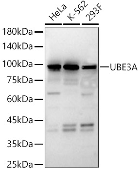
WB
Western blot analysis of various lysates, using UBE3A Rabbit pAb at 1:500 dilution. Secondary antibody: HRP Goat Anti-Rabbit IgG (H+L) at 1:10000 dilution. Lysates/proteins: 25μg per lane. Blocking buffer: 3% nonfat dry milk in TBST. Detection: ECL Basic Kit. Exposure time: 60s.
IHC
Immunohistochemistry analysis of paraffin embedded human liver cancer using UBE3A Rabbit pAb at dilution of 1:100 (40x lens).Perform high pressure antigen retrieval with 10 mM citrate buffer pH 6.0 before commencing with IHC staining protocol.
ICC/IF
Immunofluorescence analysis of HeLa cells using UBE3A Rabbit pAb at dilution of 1:50 (40x lens). Secondary antibody: Cy3 Goat Anti-Rabbit IgG (H+L)at 1:500 dilution. Blue: DAPI for nuclear staining.| Application Notes | WB:1:100-1:500 IHC:1:50-1:200 ICC/IF:1:50-1:200 |
|---|
| Form | Liquid |
|---|---|
| Storage Instructions | Store at -20℃. Avoid freeze / thaw cycles. |
| Storage Buffer | Buffer: PBS with 0.01% thimerosal, 50% glycerol, pH7.3. |
抱歉,暂无相关文献







