| 存储条件 |
|---|
| Product Name | Chk1 Recombinant Rabbit Monoclonal Antibody |
|---|---|
| Antibody Type | Primary Antibodies |
| Immunogen | Recombinant protein within |
| Clonality | monoclonal |
|---|---|
| Isotype | IgG |
| Host Species | Rabbit |
| Tested Applications | ICC/IFIPWB |
| WB:1:1000 ICC/IF:1:50 IP:1-2μg/sample | |
| Species Reactivity | HumanMouseRat |
| Concentration | 1mg/ml |
| Purification | Protein A |
| Gene Symbol | CHEK1 |
|---|---|
| Gene Synonyms | CHK1 OZEMA21 |
| Gene Full Name | checkpoint kinase 1 |
| Gene Summary | The protein encoded by this gene belongs to the Ser/Thr protein kinase family. It is required for checkpoint mediated cell cycle arrest in response to DNA damage or the presence of unreplicated DNA. This protein acts to integrate signals from ATM and ATR, two cell cycle proteins involved in DNA damage responses, that also associate with chromatin in meiotic prophase I. Phosphorylation of CDC25A protein phosphatase by this protein is required for cells to delay cell cycle progression in response to double-strand DNA breaks. Several alternatively spliced transcript variants have been found for this gene. [provided by RefSeq, Oct 2011] |
| Alternative Names | C85740 antibody Cell cycle checkpoint kinase antibody Checkpoint S. pombe homolog of 1 antibody Checkpoint kinase 1 antibody Checkpoint kinase 1 homolog (S. pombe) antibody CHEK 1 antibody Chek1 antibody Chk 1 antibody Chk1 antibody CHK1 checkpoint homolog (S. pombe) antibody C85740 antibody Cell cycle checkpoint kinase antibody Checkpoint S. pombe homolog of 1 antibody Checkpoint kinase 1 antibody Checkpoint kinase 1 homolog (S. pombe) antibody CHEK 1 antibody Chek1 antibody Chk 1 antibody Chk1 antibody CHK1 checkpoint homolog (S. pombe) antibody CHK1_HUMAN antibody EC 2.7.11.1 antibody rad27 antibody Serine/threonine protein kinase Chk1 antibody Serine/threonine-protein kinase CHK1 antibody STT3 subunit of the oligosaccharyltransferase complex homolog A (S. cerevisiae) antibody |
| Molecular Weight(MW) | 54kDa |
| Cellular Localization | Nucleus, Chromosome, Cytoplasm, cytoskeleton, microtubule organizing center, centrosome. |
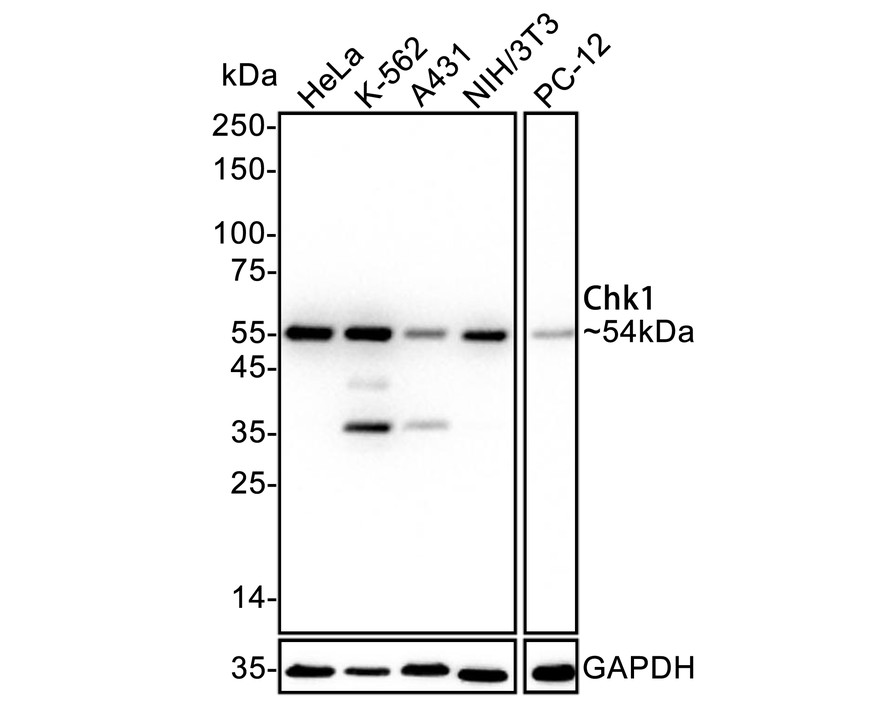
WB
Western blot analysis of Chk1 on different lysates with Rabbit anti-Chk1 antibody at 1/1,000 dilution. Lane 1: HeLa cell lysate, Lane 2: K-562 cell lysate, Lane 3: A431 cell lysate, Lane 4: NIH/3T3 cell lysate, Lane 5: PC-12 cell lysate, Lysates/proteins at 20 µg/Lane. Exposure time: 25 seconds; 4-20% SDS-PAGE gel. Proteins were transferred to a PVDF membrane and blocked with 5% NFDM/TBST for 1 hour at room temperature. The primary antibody at 1/1,000 dilution was used in 5% NFDM/TBST at 4℃ overnight. Goat Anti-Rabbit IgG - HRP Secondary Antibody at 1/50,000 dilution was used for 1 hour at room temperature.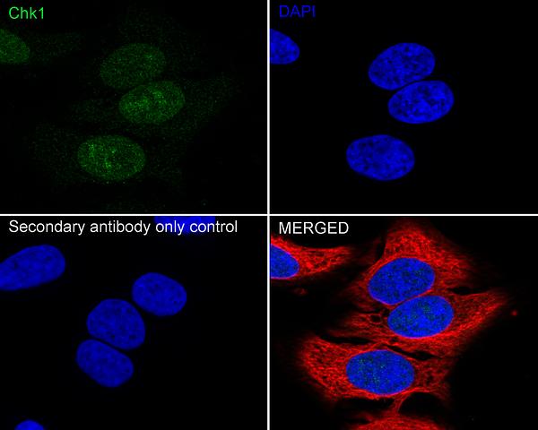
ICC/IF
Immunocytochemistry analysis of HeLa cells labeling Chk1 with Rabbit anti-Chk1 antibody at 1/50 dilution. Cells were fixed in 4% paraformaldehyde for 15 minutes at room temperature, permeabilized with 0.1% Triton X-100 in PBS for 5 minutes at room temperature, then blocked with 1% BSA in 10% negative goat serum for 1 hour at room temperature. Cells were then incubated with Rabbit anti-Chk1 antibody at 1/50 dilution in 1% BSA in PBST overnight at 4 ℃. Goat Anti-Rabbit IgG H&L (488) was used as the secondary antibody at 1/1,000 dilution. PBS instead of the primary antibody was used as the secondary antibody only control. Nuclear DNA was labelled in blue with DAPI. Beta tubulin (red) was stained at 1/100 dilution overnight at +4℃. Goat Anti-Mouse IgG H&L (594) was used as the secondary antibody at 1/1,000 dilution.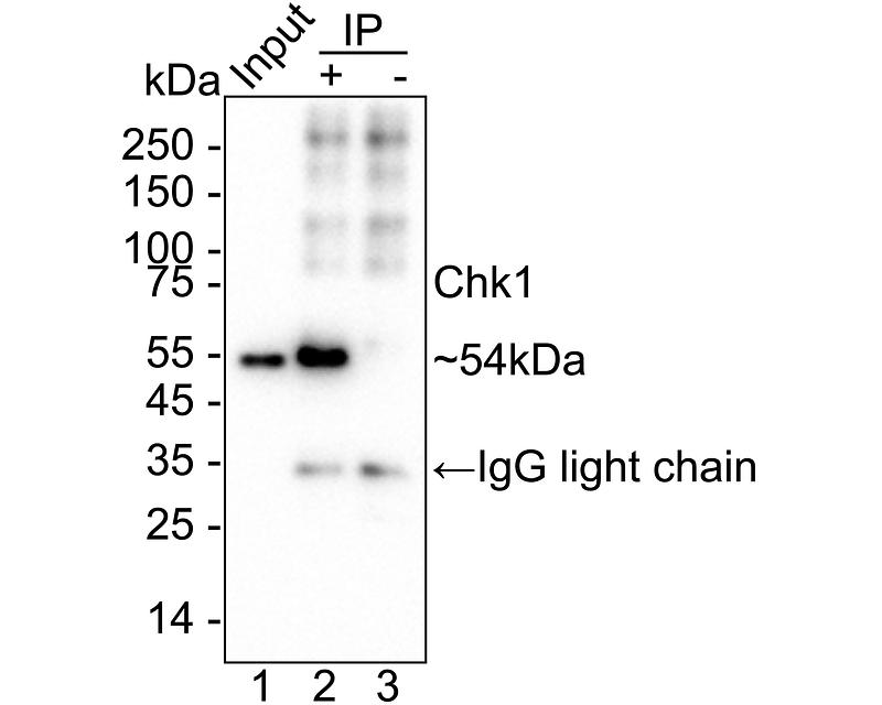
IP
Chk1 was immunoprecipitated from 0.2 mg HeLa cell lysate with Rabbit anti-Chk1 antibody at 2 µg/25 µl agarose. Western blot was performed from the immunoprecipitate using Rabbit anti-Chk1 antibody at 1/1,000 dilution. Anti-Rabbit IgG for IP Nano-secondary antibody at 1/5,000 dilution was used for 1 hour at room temperature. Lane 1: HeLa cell lysate (input), Lane 2: Rabbit anti-Chk1 antibody IP in HeLa cell lysate, Lane 3: Rabbit IgG instead of Rabbit anti-Chk1 antibody in HeLa cell lysate. Blocking/Dilution buffer: 5% NFDM/TBST. Exposure time: 20 seconds.| Application Notes | WB:1:1000 ICC/IF:1:50 IP:1-2μg/sample |
|---|
| Form | Liquid |
|---|---|
| Storage Instructions | Store at +4℃ after thawing. Aliquot store at -20℃. Avoid repeated freeze / thaw cycles. |
| Storage Buffer | 1*TBS (pH7.4), 0.05% BSA, 40% Glycerol. Preservative: 0.05% Sodium Azide. |
抱歉,暂无相关文献







